Structural mechanism underlying PHO1;H1-mediated phosphate transport in Arabidopsis.
Fang, S., Yang, Y., Zhang, X., Yang, Z., Zhang, M., Zhao, Y., Zhang, C., Yu, F., Wang, Y.F., Zhang, P.(2025) Nat Plants 11: 309-320
- PubMed: 39838070 Search on PubMedSearch on PubMed Central
- DOI: https://doi.org/10.1038/s41477-024-01895-6
- Primary Citation Related Structures:
9IK4, 9JF8 - PubMed Abstract:
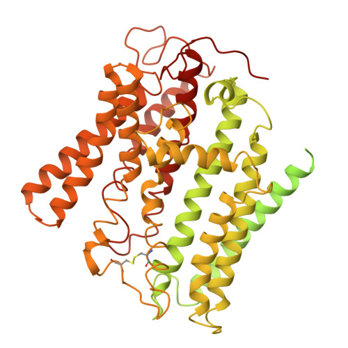
Arabidopsis PHOSPHATE 1 (AtPHO1) and its closest homologue AtPHO1;H1 are phosphate transporters that load phosphate into the xylem vessel for root-to-shoot translocation. AtPHO1 and AtPHO1;H1 are prototypical members of the unique SPX-EXS family, whose structural and molecular mechanisms remain elusive. In this study, we determined the cryogenic electron microscopy structure of AtPHO1;H1 binding with inorganic phosphate (Pi) and inositol hexakisphosphate in a closed conformation. Further molecular dynamic simulation and AlphaFold prediction support an open conformation. AtPHO1;H1 forms a domain-swapped homodimer that involves both the transmembrane ERD1/XPR1/SYG1 (EXS) domain and the cytoplasmic SYG1/Pho81/XPR1 (SPX) domain. The EXS domain presented by the SPX-EXS family represents a novel protein fold, and an independent substrate transport pathway and substrate-binding site are present in each EXS domain. Two gating residues, Trp719 and Tyr610, are identified above the substrate-binding site to control opening and closing of the pathway. The SPX domain features positively charged patches and/or residues at the dimer interface to accommodate inositol hexakisphosphate molecules, whose binding mediates dimerization and enhances AtPHO1;H1 activity. In addition, a C-terminal tail is required for AtPHO1;H1 activity. On the basis of structural and functional analysis, a working model for Pi efflux mediated by AtPHO1;H1 and its homologues was postulated, suggesting a channel-like mechanism. This study not only reveals the molecular and regulatory mechanism underlying Pi transport mediated by the unique SPX-EXS family, but also provides potential for crop engineering to enhance phosphorus-use efficiency in sustainable agriculture.
- National Key Laboratory of Plant Molecular Genetics, CAS Center for Excellence in Molecular Plant Sciences, Institute of Plant Physiology and Ecology, Chinese Academy of Sciences, Shanghai, China.
Organizational Affiliation: