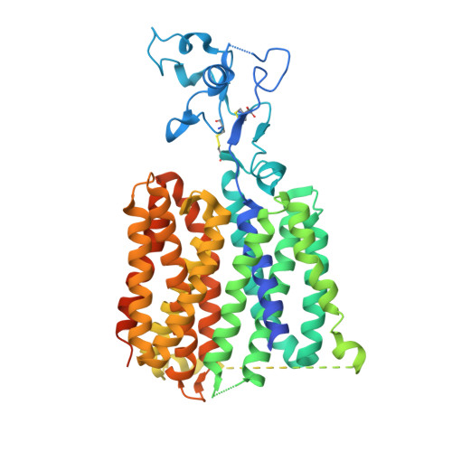
Gout, a common and painful disease, stems from hyperuricemia, where elevated blood urate levels lead to urate crystal formation in joints and kidneys. The human urate transporter 1 (hURAT1) plays a critical role in urate homeostasis by facilitating urate reabsorption in the renal proximal tubule, making it a key target for gout therapy. Pharmacological inhibition of hURAT1 with drugs such as dotinurad, benzbromarone, lesinurad, and verinurad promotes urate excretion and alleviates gout symptoms. Here, we present cryo-electron microscopy structures of native hURAT1 bound with these anti-gout drugs in the inward-open state, and with urate in inward-open, outward-open, and occluded states. Complemented by mutagenesis and cell-based assays, these structures reveal the mechanisms of urate reabsorption and hURAT1 inhibition. Our findings elucidate the molecular basis of urate transport and anti-gout medication action and provide a structural framework for the rational design of next-generation therapies for hyperuricemia and gout.
Organizational Affiliation:
Research Center for Medicinal Structural Biology, National Research Center for Translational Medicine at Shanghai, State Key Laboratory of Medical Genomics, Ruijin Hospital, Shanghai Jiao Tong University School of Medicine, Shanghai, China. wcr13215@rjh.com.cn.
State Key Laboratory of Drug Research, Shanghai Institute of Materia Medica, Chinese Academy of Sciences, Shanghai, China. wcr13215@rjh.com.cn.
School of Pharmacy, Fudan University, Shanghai, China.
State Key Laboratory of Chemical Biology, Shanghai Institute of Materia Medica, Chinese Academy of Sciences, Shanghai, China.
Lingang laboratory, Shanghai, China.
Shanghai Institute of Materia Medica, Chinese Academy of Sciences, Shanghai, China.
Division of Cardiology, Department of Internal Medicine and Hubei Key Laboratory of Genetics and Molecular Mechanism of Cardiologic Disorders, Tongji Hospital, Tongji Medical College, Huazhong University of Science and Technology, Wuhan, Hubei, China.
The National Center for Drug Screening, Shanghai Institute of Materia Medica, Chinese Academy of Sciences, Shanghai, China.
State Key Laboratory of Drug Research, Shanghai Institute of Materia Medica, Chinese Academy of Sciences, Shanghai, China.
Research Center for Medicinal Structural Biology, National Research Center for Translational Medicine at Shanghai, State Key Laboratory of Medical Genomics, Ruijin Hospital, Shanghai Jiao Tong University School of Medicine, Shanghai, China.
Research Center for Deepsea Bioresources, Sanya, Hainan, China.
Department of Pharmacology, School of Basic Medical Sciences, Fudan University, Shanghai, China.
Department of Chemistry, School of Science, The University of Tokyo, Tokyo, Japan.
Shanghai Institute of Materia Medica, Chinese Academy of Sciences, Shanghai, China. yjiang@lglab.ac.cn.
State Key Laboratory of Chemical Biology, Shanghai Institute of Materia Medica, Chinese Academy of Sciences, Shanghai, China. dhyang@simm.ac.cn.
The National Center for Drug Screening, Shanghai Institute of Materia Medica, Chinese Academy of Sciences, Shanghai, China. dhyang@simm.ac.cn.
School of Pharmacy, Hainan Medical University, Haikou, Hainan, China. dhyang@simm.ac.cn.
Research Center for Medicinal Structural Biology, National Research Center for Translational Medicine at Shanghai, State Key Laboratory of Medical Genomics, Ruijin Hospital, Shanghai Jiao Tong University School of Medicine, Shanghai, China. eric.xu@simm.ac.cn.
State Key Laboratory of Drug Research, Shanghai Institute of Materia Medica, Chinese Academy of Sciences, Shanghai, China. eric.xu@simm.ac.cn.
University of Chinese Academy of Sciences, Beijing, China. eric.xu@simm.ac.cn.
Center for Drug Research and Evaluation, National Infrastructures for Translational Medicine, Institute of Clinical Medicine, Peking Union Medical College Hospital, Beijing, China. eric.xu@simm.ac.cn.