


Crystal structure of the DH domain of human FGD6
Liu, Z.C., Chuan, J.L., Wang, G.G.To be published.
Experimental Data Snapshot
Starting Model: experimental
View more details
wwPDB Validation 3D Report Full Report

Entity ID: 1 | |||||
|---|---|---|---|---|---|
| Molecule | Chains | Sequence Length | Organism | Details | Image |
| FYVE, RhoGEF and PH domain-containing protein 6 | 189 | Homo sapiens | Mutation(s): 0 Gene Names: FGD6, KIAA1362, ZFYVE24 | 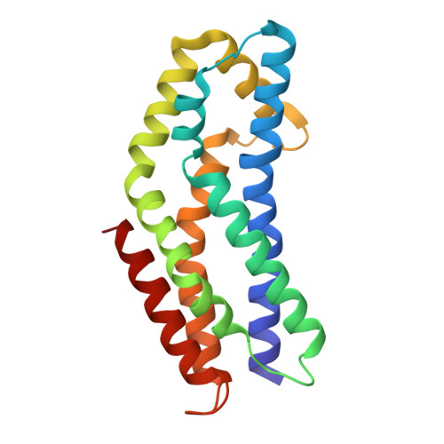 | |
UniProt & NIH Common Fund Data Resources | |||||
PHAROS: Q6ZV73 GTEx: ENSG00000180263 | |||||
Entity Groups | |||||
| Sequence Clusters | 30% Identity50% Identity70% Identity90% Identity95% Identity100% Identity | ||||
| UniProt Group | Q6ZV73 | ||||
Sequence AnnotationsExpand | |||||
Reference Sequence | |||||
| Length ( Å ) | Angle ( ˚ ) |
|---|---|
| a = 74.197 | α = 90 |
| b = 74.197 | β = 90 |
| c = 40.398 | γ = 90 |
| Software Name | Purpose |
|---|---|
| PHENIX | refinement |
| autoPROC | data reduction |
| xia2 | data scaling |
| PHASER | phasing |
| Funding Organization | Location | Grant Number |
|---|---|---|
| Not funded | -- |


