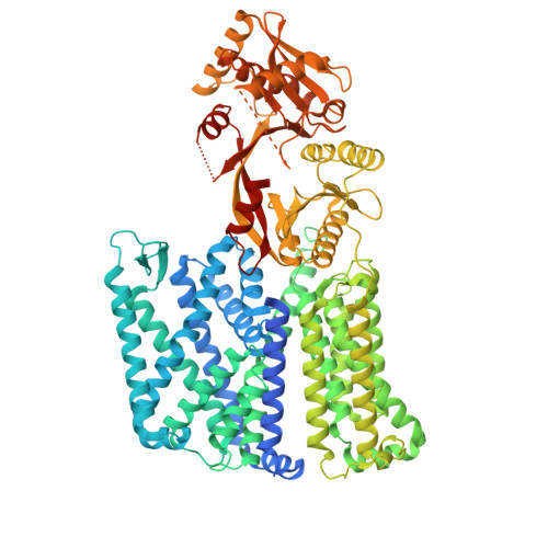
Cryo-EM structures of multiple-peptide resistance factor (MprF) from Pseudomonas aeruginosa.
Jha, S., Vinothkumar, K.R.(2026) FEBS J
- PubMed: 42175596 Search on PubMed
- DOI: https://doi.org/10.1111/febs.70519
- Primary Citation Related Structures:
9J8Q, 9J8R, 9J8S, 9W50, 9W51 - PubMed Abstract:
Aminoacylation of the lipid head group in many bacteria is carried out by bi-functional enzymes called MprF, which encode a soluble synthase domain that typically transfers lysine or alanine from a tRNA to lipid head groups. The modified lipid is subsequently translocated across the leaflets by a transmembrane domain. This modification of lipids probably evolved to adapt to the environment where the microbes reside. Here, we describe the cryo-EM structures of MprF enzyme from Pseudomonas aeruginosa, revealing a dimeric enzyme with a distinct architecture when compared with the homologous Rhizobium enzymes, and validate this arrangement with biochemical analyses. The cryo-EM maps and the models in detergent micelle and nanodisc reveal a conformational change of the terminal helix of the synthase domain, highlighting the dynamic elements in the enzyme that might facilitate catalysis. Several lipid-like densities are observed in the cryo-EM maps, which might indicate the path taken by the lipids, coupling the function of the two domains. The structures allow postulation of the binding modes of tRNA and lipid transport, and suggest that the mobile secondary structural elements in the synthase domain might play a mechanistic role in these functions.
- National Centre for Biological Sciences, Tata Institute of Fundamental Research, Bengaluru, India.
Organizational Affiliation: