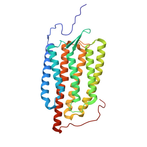
Cryo-EM structure of a blue-shifted channelrhodopsin from Klebsormidium nitens.
Wang, Y.Z., Natsume, K., Tanaka, T., Hososhima, S., Tashiro, R., Sano, F.K., Akasaka, H., Tsunoda, S.P., Shihoya, W., Kandori, H., Nureki, O.(2025) Nat Commun 16: 5297-5297
- PubMed: 40533461 Search on PubMedSearch on PubMed Central
- DOI: https://doi.org/10.1038/s41467-025-59299-9
- Primary Citation Related Structures:
9J7W - PubMed Abstract:
Channelrhodopsins (ChRs) are light-gated ion channels and invaluable tools for optogenetic applications. Recent developments in multicolor optogenetics, in which different neurons are controlled by multiple colors of light simultaneously, have increased the demand for ChR mutants with more distant absorption wavelengths. Here we report the 2.7 Å-resolution cryo-electron microscopy structure of a ChR from Klebsormidium nitens (KnChR), which is one of the most blue-shifted ChRs. The structure elucidates the 6-s-cis configuration of the retinal chromophore, indicating its contribution to a distinctive blue shift in action spectra. The unique architecture of the C-terminal region reveals its role in the allosteric modulation of channel kinetics, enhancing our understanding of its functional dynamics. Employing a rational approach, we developed mutants with blue-shifted action spectra. Finally, we confirm that UV or deep-blue light can activate KnChR-transfected precultured neurons, expanding its utility in optogenetic applications. Our findings contribute valuable insights to advance optogenetic tools and enable refined capabilities in neuroscience experiments.
- Department of Biological Sciences, Graduate School of Science, The University of Tokyo, Tokyo, Japan.
Organizational Affiliation: