9J6N | pdb_00009j6n

beta-D-galactofuranosidase ORF1110 from Streptomyces sp. JHA19, D-iminogalactitol complex


Experimental Data Snapshot

  • Method: X-RAY DIFFRACTION
  • Resolution: 1.70 Å
  • R-Value Free: 
    0.189 (Depositor), 0.201 (DCC) 
  • R-Value Work: 
    0.153 (Depositor), 0.167 (DCC) 
  • R-Value Observed: 
    0.155 (Depositor) 

wwPDB Validation   3D Report Full Report


Ligand Structure Quality Assessment 


This is version 1.1 of the entry. See complete history


Literature

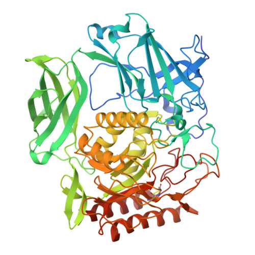
Crystal structure of beta-d-galactofuranosidase from Streptomyces sp. JHA19 in complex with an inhibitor provides insights into substrate specificity.

Fujio, N.Yamada, C.Kashima, T.Matsunaga, E.Nash, R.J.Takegawa, K.Fushinobu, S.

(2024) FEBS Lett 598: 2866-2875

  • DOI: https://doi.org/10.1002/1873-3468.15056
  • Primary Citation of Related Structures:  
    9J6M, 9J6N

  • PubMed Abstract: 

    d-Galactofuranose (Galf) is widely distributed in glycoconjugates of pathogenic microbes. β-d-Galactofuranosidase (Galf-ase) from Streptomyces sp. JHA19 (ORF1110) belongs to glycoside hydrolase (GH) family 2 and is the first identified Galf-specific degradation enzyme. Here, the crystal structure of ORF1110 in complex with a mechanism-based potent inhibitor, d-iminogalactitol (K i  = 65 μm) was solved. ORF1110 binds to the C5-C6 hydroxy groups of d-iminogalactitol with an extensive and integral hydrogen bond network, a key interaction that discriminates the substrates. The active site structure of ORF1110 is largely different from those of β-glucuronidases and β-galactosidases in the same GH2 family. A C-terminal domain of ORF1110 is predicted to be a carbohydrate-binding module family 42 that may bind Galf. The structural insights into Galf-ase will contribute to the investigation of therapeutic tools against pathogens.


  • Organizational Affiliation
    • Department of Biotechnology, The University of Tokyo, Japan.

Macromolecules
Find similar proteins by:  (by identity cutoff)  |  3D Structure
Entity ID: 1
MoleculeChains Sequence LengthOrganismDetailsImage
Beta-D-galactofuranosidase 2
A, B
612Streptomyces sp. JHA19Mutation(s): 0 
Entity Groups  
Sequence Clusters30% Identity50% Identity70% Identity90% Identity95% Identity100% Identity
Sequence Annotations
Expand
  • Reference Sequence
Small Molecules
Ligands 5 Unique
IDChains Name / Formula / InChI Key2D Diagram3D Interactions
P6G
Query on P6G

Download Ideal Coordinates CCD File 
C [auth A]HEXAETHYLENE GLYCOL
C12 H26 O7
IIRDTKBZINWQAW-UHFFFAOYSA-N
PG4
Query on PG4

Download Ideal Coordinates CCD File 
H [auth B]TETRAETHYLENE GLYCOL
C8 H18 O5
UWHCKJMYHZGTIT-UHFFFAOYSA-N
A1L3Z (Subject of Investigation/LOI)
Query on A1L3Z

Download Ideal Coordinates CCD File 
F [auth A],
J [auth B]
(2~{S},3~{S},4~{S})-2-[(1~{S})-1,2-bis(oxidanyl)ethyl]pyrrolidine-3,4-diol
C6 H13 N O4
RVNSAAIWCWTCTJ-BGPJRJDNSA-N
MPD
Query on MPD

Download Ideal Coordinates CCD File 
D [auth A],
E [auth A],
I [auth B]
(4S)-2-METHYL-2,4-PENTANEDIOL
C6 H14 O2
SVTBMSDMJJWYQN-YFKPBYRVSA-N
MG
Query on MG

Download Ideal Coordinates CCD File 
G [auth A],
K [auth B]
MAGNESIUM ION
Mg
JLVVSXFLKOJNIY-UHFFFAOYSA-N
Experimental Data & Validation

Experimental Data

  • Method: X-RAY DIFFRACTION
  • Resolution: 1.70 Å
  • R-Value Free:  0.189 (Depositor), 0.201 (DCC) 
  • R-Value Work:  0.153 (Depositor), 0.167 (DCC) 
  • R-Value Observed: 0.155 (Depositor) 
Space Group: P 1 21 1
Unit Cell:
Length ( Å )Angle ( ˚ )
a = 82.364α = 90
b = 57.346β = 94.93
c = 109.732γ = 90
Software Package:
Software NamePurpose
REFMACrefinement
XDSdata reduction
Aimlessdata scaling
CRANK2phasing

Structure Validation

View Full Validation Report


Ligand Structure Quality Assessment 


Entry History & Funding Information

Deposition Data


Funding OrganizationLocationGrant Number
Not funded--

Revision History  (Full details and data files)

  • Version 1.0: 2024-11-27
    Type: Initial release
  • Version 1.1: 2024-12-25
    Changes: Database references