9J6K | pdb_00009j6k

HBx complexed with DDB1


Experimental Data Snapshot

  • Method: ELECTRON MICROSCOPY
  • Resolution: 2.68 Å
  • Aggregation State: PARTICLE 
  • Reconstruction Method: SINGLE PARTICLE 

wwPDB Validation   3D Report Full Report


This is version 1.1 of the entry. See complete history


Literature

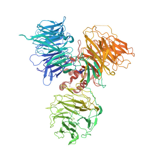
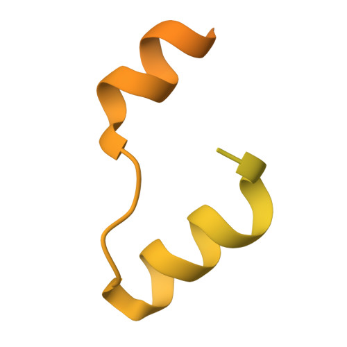
Structural basis of the hepatitis B virus X protein in complex with DDB1.

Tanaka, H.Diogo Dias, J.Jay, B.Kita, S.Sasaki, M.Takeda, H.Kishimoto, N.Sasaki, S.Misumi, S.Mizokami, M.Neuveut, C.Sumikama, T.Shibata, M.Maenaka, K.Machida, S.

(2025) Proc Natl Acad Sci U S A 122: e2421325122-e2421325122

  • DOI: https://doi.org/10.1073/pnas.2421325122
  • Primary Citation of Related Structures:  
    9J6J, 9J6K

  • PubMed Abstract: 

    A cure for chronic hepatitis B requires eliminating or permanently silencing covalently closed circular DNA (cccDNA). A pivotal target of this approach is the hepatitis B virus (HBV) X protein (HBx), which is a key factor that promotes transcription from cccDNA. However, the HBx structure remains unsolved. Here, we present the cryoelectron microscopy structure of HBx in complex with DDB1, which is an essential complex for cccDNA transcription. In this structure, hydrophobic interactions within HBx were identified, and mutational analysis highlighted their importance in the HBV life cycle. Our biochemical analysis revealed that the HBx-DDB1 complex directly interacts simultaneously with NSE3, which is a component of the SMC5/6 complex, and Spindlin1. Additionally, HBx-DDB1 complex dynamics were explored via high-speed atomic force microscopy. These findings provide comprehensive insights into the structure and function of HBx in HBV replication.


  • Organizational Affiliation
    • Department of Structural Virology, National Institute of Global Health and Medicine, Japan Institute for Health Security, Shinjuku-ku, Tokyo 162-8655, Japan.

Macromolecules
Find similar proteins by:  (by identity cutoff)  |  3D Structure
Entity ID: 1
MoleculeChains Sequence LengthOrganismDetailsImage
DNA damage-binding protein 11,144Homo sapiensMutation(s): 0 
Gene Names: DDB1XAP1
UniProt & NIH Common Fund Data Resources
Find proteins for Q16531 (Homo sapiens)
Explore Q16531 
Go to UniProtKB:  Q16531
PHAROS:  Q16531
GTEx:  ENSG00000167986 
Entity Groups  
Sequence Clusters30% Identity50% Identity70% Identity90% Identity95% Identity100% Identity
UniProt GroupQ16531
Sequence Annotations
Expand
  • Reference Sequence
Find similar proteins by:  (by identity cutoff)  |  3D Structure
Entity ID: 2
MoleculeChains Sequence LengthOrganismDetailsImage
Protein X171Hepatitis B virusMutation(s): 0 
UniProt
Find proteins for Q77UW5 (Hepatitis B virus)
Explore Q77UW5 
Go to UniProtKB:  Q77UW5
Entity Groups  
Sequence Clusters30% Identity50% Identity70% Identity90% Identity95% Identity100% Identity
UniProt GroupQ77UW5
Sequence Annotations
Expand
  • Reference Sequence
Experimental Data & Validation

Experimental Data

  • Method: ELECTRON MICROSCOPY
  • Resolution: 2.68 Å
  • Aggregation State: PARTICLE 
  • Reconstruction Method: SINGLE PARTICLE 
EM Software:
TaskSoftware PackageVersion
MODEL REFINEMENTPHENIX

Structure Validation

View Full Validation Report


Entry History & Funding Information

Deposition Data


Funding OrganizationLocationGrant Number
Other governmentJapan22T003
Other governmentJapan23A1017
Japan Agency for Medical Research and Development (AMED)JapanJP20ae0101047
Japan Agency for Medical Research and Development (AMED)JapanJP22ama121037
Japan Agency for Medical Research and Development (AMED)JapanJP223fa627005

Revision History  (Full details and data files)

  • Version 1.0: 2025-06-04
    Type: Initial release
  • Version 1.1: 2025-06-25
    Changes: Data collection, Database references