9J19 | pdb_00009j19

The crystal structure of COVID-19 main protease in complex with an inhibitor minocycline


Experimental Data Snapshot

  • Method: X-RAY DIFFRACTION
  • Resolution: 2.70 Å
  • R-Value Free: 
    0.314 (Depositor), 0.325 (DCC) 
  • R-Value Work: 
    0.250 (Depositor), 0.259 (DCC) 
  • R-Value Observed: 
    0.253 (Depositor) 

Starting Model: experimental
View more details

wwPDB Validation   3D Report Full Report


Ligand Structure Quality Assessment 


This is version 1.2 of the entry. See complete history


Literature

Structural and Mechanistic Insights into the Main Protease (Mpro) Dimer Interface Destabilization Inhibitor: Unveiling New Therapeutic Avenues against SARS-CoV-2.

Singh, A.Jangid, K.Nehul, S.Dhaka, P.Rani, R.Pareek, A.Sharma, G.K.Kumar, P.Tomar, S.

(2025) Biochemistry 64: 1589-1605

  • DOI: https://doi.org/10.1021/acs.biochem.4c00535
  • Primary Citation of Related Structures:  
    9J19

  • PubMed Abstract: 

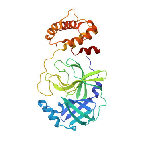
    SARS-CoV-2 variant recurrence has emphasized the imperative prerequisite for effective antivirals. The main protease (Mpro) of SARS-CoV-2 is crucial for viral replication, making it one of the prime and promising antiviral targets. Mpro features several druggable sites, including active sites and allosteric sites near the dimerization interface, that regulate its catalytic activity. This study identified six highly efficacious antiviral SARS-CoV-2 compounds (WIN-62577, KT185, bexarotene, ledipasvir, diacerein, and simepervir) using structure-based virtual screening of compound libraries against Mpro. Using SPR and ITC, the binding of selected inhibitory compounds to the target Mpro was validated. The FRET-based protease assay demonstrated that the identified molecules effectively inhibit Mpro with IC 50 values in the range from 0.64 to 11.98 μM. Additionally, in vitro cell-based antiviral assays showed high efficacy with EC 50 values in the range of 1.51 to 18.92 μM. The crystal structure of the Mpro-minocycline complex detailed the possible inhibition mechanism of minocycline, an FDA-approved antibiotic. Minocycline binds to an allosteric site, revealing residues critical for the loss of protease activity due to destabilization of molecular interactions at the dimeric interface, which are crucial for the proteolytic activity of Mpro. The study suggests that the binding of minocycline to the allosteric site may play a role in Mpro dimer destabilization and direct the rational design of minocycline derivatives as antiviral drugs.


  • Organizational Affiliation
    • Department of Biosciences and Bioengineering, Indian Institute of Technology, Roorkee, Uttarakhand 247667, India.

Macromolecules
Find similar proteins by:  (by identity cutoff)  |  3D Structure
Entity ID: 1
MoleculeChains Sequence LengthOrganismDetailsImage
3C-like proteinase nsp5
A, B
296Severe acute respiratory syndrome coronavirus 2Mutation(s): 0 
Gene Names: rep1a-1b
EC: 3.4.22.69
UniProt
Find proteins for P0DTD1 (Severe acute respiratory syndrome coronavirus 2)
Explore P0DTD1 
Go to UniProtKB:  P0DTD1
Entity Groups  
Sequence Clusters30% Identity50% Identity70% Identity90% Identity95% Identity100% Identity
UniProt GroupP0DTD1
Sequence Annotations
Expand
  • Reference Sequence
Small Molecules
Ligands 1 Unique
IDChains Name / Formula / InChI Key2D Diagram3D Interactions
MIY (Subject of Investigation/LOI)
Query on MIY

Download Ideal Coordinates CCD File 
C [auth A](4S,4AS,5AR,12AS)-4,7-BIS(DIMETHYLAMINO)-3,10,12,12A-TETRAHYDROXY-1,11-DIOXO-1,4,4A,5,5A,6,11,12A-OCTAHYDROTETRACENE-2- CARBOXAMIDE
C23 H27 N3 O7
DYKFCLLONBREIL-KVUCHLLUSA-N
Experimental Data & Validation

Experimental Data

  • Method: X-RAY DIFFRACTION
  • Resolution: 2.70 Å
  • R-Value Free:  0.314 (Depositor), 0.325 (DCC) 
  • R-Value Work:  0.250 (Depositor), 0.259 (DCC) 
  • R-Value Observed: 0.253 (Depositor) 
Space Group: P 21 21 21
Unit Cell:
Length ( Å )Angle ( ˚ )
a = 67.945α = 90
b = 90.763β = 90
c = 101.307γ = 90
Software Package:
Software NamePurpose
REFMACrefinement
CrysalisProdata reduction
Aimlessdata scaling
MOLREPphasing

Structure Validation

View Full Validation Report


Ligand Structure Quality Assessment 


Entry History & Funding Information

Deposition Data


Funding OrganizationLocationGrant Number
Science and Engineering Research Board (SERB)IndiaIPA/2020/000054

Revision History  (Full details and data files)

  • Version 1.0: 2025-02-12
    Type: Initial release
  • Version 1.1: 2025-03-12
    Changes: Structure summary
  • Version 1.2: 2025-04-09
    Changes: Database references