


Crystal structure of RhoA-TP1001 complex
Zhu, L., Li, H., Chang, L., Hu, X.To be published.
Experimental Data Snapshot
Starting Model: experimental
View more details
Entity ID: 1 | |||||
|---|---|---|---|---|---|
| Molecule | Chains | Sequence Length | Organism | Details | Image |
| Transforming protein RhoA | 193 | Homo sapiens | Mutation(s): 0 Gene Names: RHOA, ARH12, ARHA, RHO12 EC: 3.6.5.2 | 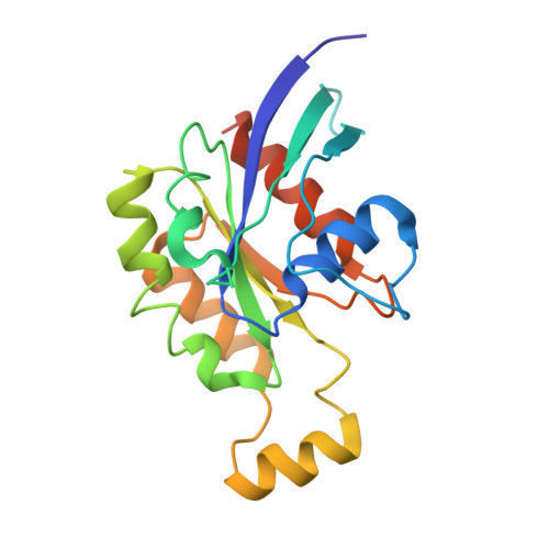 | |
UniProt & NIH Common Fund Data Resources | |||||
PHAROS: P61586 GTEx: ENSG00000067560 | |||||
Entity Groups | |||||
| Sequence Clusters | 30% Identity50% Identity70% Identity90% Identity95% Identity100% Identity | ||||
| UniProt Group | P61586 | ||||
Sequence AnnotationsExpand | |||||
Reference Sequence | |||||
| Ligands 4 Unique | |||||
|---|---|---|---|---|---|
| ID | Chains | Name / Formula / InChI Key | 2D Diagram | 3D Interactions | |
| GDP Download:Ideal Coordinates CCD File | E [auth A], G [auth B], J [auth C], M [auth D] | GUANOSINE-5'-DIPHOSPHATE C10 H15 N5 O11 P2 QGWNDRXFNXRZMB-UUOKFMHZSA-N |  | ||
| EPE Download:Ideal Coordinates CCD File | P [auth D] | 4-(2-HYDROXYETHYL)-1-PIPERAZINE ETHANESULFONIC ACID C8 H18 N2 O4 S JKMHFZQWWAIEOD-UHFFFAOYSA-N |  | ||
| U6L (Subject of Investigation/LOI) Download:Ideal Coordinates CCD File | L [auth D] | (1~{R})-1-(3-ethylphenyl)ethane-1,2-diol C10 H14 O2 ZPNNJYNIHQZIJX-JTQLQIEISA-N |  | ||
| MG Download:Ideal Coordinates CCD File | F [auth A] H [auth B] I [auth B] K [auth C] N [auth D] | MAGNESIUM ION Mg JLVVSXFLKOJNIY-UHFFFAOYSA-N |  | ||
| Length ( Å ) | Angle ( ˚ ) |
|---|---|
| a = 81.24 | α = 90 |
| b = 124.8 | β = 90 |
| c = 173.5 | γ = 90 |
| Software Name | Purpose |
|---|---|
| REFMAC | refinement |
| HKL-3000 | data scaling |
| HKL-3000 | data reduction |
| PHASER | phasing |
| Funding Organization | Location | Grant Number |
|---|---|---|
| National Natural Science Foundation of China (NSFC) | China | 82150208 |


