9IYQ | pdb_00009iyq

Structure of the human GluN1-N2B NMDA receptors in the Mg2+ free state

  • Classification: MEMBRANE PROTEIN
  • Organism(s): Homo sapiens
  • Expression System: Homo sapiens
  • Mutation(s): No 
  • Membrane Protein: Yes  PDBTM

  • Deposited: 2024-07-31 Released: 2025-03-05 
  • Deposition Author(s): Huang, X., Sun, X., Zhu, S.
  • Funding Organization(s): Ministry of Science and Technology (MoST, China), National Natural Science Foundation of China (NSFC), Chinese Academy of Sciences

Experimental Data Snapshot

  • Method: ELECTRON MICROSCOPY
  • Resolution: 3.18 Å
  • Aggregation State: CELL 
  • Reconstruction Method: SINGLE PARTICLE 

wwPDB Validation   3D Report Full Report


This is version 1.3 of the entry. See complete history


Literature

Structural insights into the diverse actions of magnesium on NMDA receptors.

Huang, X.Sun, X.Wang, Q.Zhang, J.Wen, H.Chen, W.J.Zhu, S.

(2025) Neuron 113: 1006

  • DOI: https://doi.org/10.1016/j.neuron.2025.01.021
  • Primary Citation of Related Structures:  
    9IYP, 9IYQ

  • PubMed Abstract: 

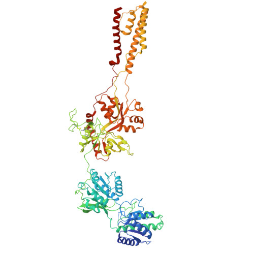
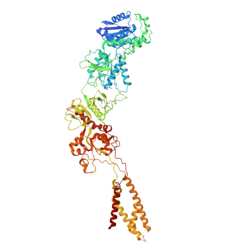
    Magnesium (Mg 2+ ) is a key regulatory ion of N-methyl-ᴅ-aspartate (NMDA) receptors, including conferring them to function as coincidence detectors for excitatory synaptic transmission. However, the structural basis underlying the Mg 2+ action on NMDA receptors remains unclear. Here, we report the cryo-EM structures of GluN1-N2B receptors and identify three distinct Mg 2+ -binding pockets. Specifically, site Ⅰ is located at the selectivity filter where an asparagine ring forms coordination bonds with Mg 2+ and is responsible for the voltage-dependent block. Sites Ⅱ and Ⅲ are located at the N-terminal domain (NTD) of the GluN2B subunit and involved in the allosteric potentiation and inhibition, respectively. Site Ⅱ consists of three acidic residues, and the combination of three mutations abolishes the GluN2B-specific Mg 2+ potentiation, while site Ⅲ overlaps with the Zn 2+ pocket, and mutations here significantly reduce the inhibition. Our study enhances the understanding of multifaceted roles of Mg 2+ in NMDA receptors and synaptic plasticity.


  • Organizational Affiliation
    • Institute of Neuroscience, CAS Center for Excellence in Brain Science and Intelligence Technology, Chinese Academy of Sciences, Shanghai 200031, China; Department of Neurology and Institute of Neurology of First Affiliated Hospital, Institute of Neuroscience, Fujian Key Laboratory of Molecular Neurology, Fujian Medical University, Fuzhou, Fujian 350005, China.

Macromolecules
Find similar proteins by:  (by identity cutoff)  |  3D Structure
Entity ID: 1
MoleculeChains Sequence LengthOrganismDetailsImage
Glutamate receptor ionotropic, NMDA 1
A, C
847Homo sapiensMutation(s): 0 
Gene Names: GRIN1NMDAR1
Membrane Entity: Yes 
UniProt & NIH Common Fund Data Resources
Find proteins for Q05586 (Homo sapiens)
Explore Q05586 
Go to UniProtKB:  Q05586
PHAROS:  Q05586
GTEx:  ENSG00000176884 
Entity Groups  
Sequence Clusters30% Identity50% Identity70% Identity90% Identity95% Identity100% Identity
UniProt GroupQ05586
Glycosylation
Glycosylation Sites: 5Go to GlyGen: Q05586-1
Sequence Annotations
Expand
  • Reference Sequence
Find similar proteins by:  (by identity cutoff)  |  3D Structure
Entity ID: 2
MoleculeChains Sequence LengthOrganismDetailsImage
Glutamate receptor ionotropic, NMDA 2B
B, D
842Homo sapiensMutation(s): 0 
Gene Names: GRIN2BNMDAR2B
Membrane Entity: Yes 
UniProt & NIH Common Fund Data Resources
Find proteins for Q13224 (Homo sapiens)
Explore Q13224 
Go to UniProtKB:  Q13224
PHAROS:  Q13224
GTEx:  ENSG00000273079 
Entity Groups  
Sequence Clusters30% Identity50% Identity70% Identity90% Identity95% Identity100% Identity
UniProt GroupQ13224
Sequence Annotations
Expand
  • Reference Sequence
Small Molecules
Ligands 3 Unique
IDChains Name / Formula / InChI Key2D Diagram3D Interactions
7RC
Query on 7RC

Download Ideal Coordinates CCD File 
I [auth B],
P [auth D]
(2R)-4-(3-phosphonopropyl)piperazine-2-carboxylic acid
C8 H17 N2 O5 P
CUVGUPIVTLGRGI-SSDOTTSWSA-N
NAG
Query on NAG

Download Ideal Coordinates CCD File 
F [auth A]
G [auth A]
H [auth A]
K [auth C]
L [auth C]
F [auth A],
G [auth A],
H [auth A],
K [auth C],
L [auth C],
M [auth C],
N [auth C],
O [auth C]
2-acetamido-2-deoxy-beta-D-glucopyranose
C8 H15 N O6
OVRNDRQMDRJTHS-FMDGEEDCSA-N
GLY
Query on GLY

Download Ideal Coordinates CCD File 
E [auth A],
J [auth C]
GLYCINE
C2 H5 N O2
DHMQDGOQFOQNFH-UHFFFAOYSA-N
Experimental Data & Validation

Experimental Data

  • Method: ELECTRON MICROSCOPY
  • Resolution: 3.18 Å
  • Aggregation State: CELL 
  • Reconstruction Method: SINGLE PARTICLE 
EM Software:
TaskSoftware PackageVersion
MODEL REFINEMENTPHENIX

Structure Validation

View Full Validation Report


Entry History & Funding Information

Deposition Data


Funding OrganizationLocationGrant Number
Ministry of Science and Technology (MoST, China)China2022ZD0212700
National Natural Science Foundation of China (NSFC)China32221003
Chinese Academy of SciencesChinato S.Z.

Revision History  (Full details and data files)

  • Version 1.0: 2025-03-05
    Type: Initial release
  • Version 1.1: 2025-03-12
    Changes: Data collection, Database references
  • Version 1.2: 2025-04-16
    Changes: Data collection, Database references
  • Version 1.3: 2025-07-23
    Changes: Data collection