


Crystal structure of Chitinase from Vibrio parahaemolyticus at pH6.5
Cheng, Q., Zhang, J.To be published.
Experimental Data Snapshot
Starting Model: in silico
View more details
wwPDB Validation 3D Report Full Report

Entity ID: 1 | |||||
|---|---|---|---|---|---|
| Molecule | Chains | Sequence Length | Organism | Details | Image |
| Glycoside hydrolase family 18 protein | 411 | Vibrio parahaemolyticus | Mutation(s): 0 Gene Names: HKB16_33210 EC: 3.2.1.14 | 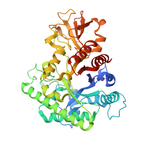 | |
UniProt | |||||
Find proteins for A0A7Y0XG39 (Vibrio parahaemolyticus) Explore A0A7Y0XG39 Go to UniProtKB: A0A7Y0XG39 | |||||
Entity Groups | |||||
| Sequence Clusters | 30% Identity50% Identity70% Identity90% Identity95% Identity100% Identity | ||||
| UniProt Group | A0A7Y0XG39 | ||||
Sequence AnnotationsExpand | |||||
Reference Sequence | |||||
| Length ( Å ) | Angle ( ˚ ) |
|---|---|
| a = 123.06 | α = 90 |
| b = 50.42 | β = 126.53 |
| c = 95.33 | γ = 90 |
| Software Name | Purpose |
|---|---|
| REFMAC | refinement |
| XDS | data reduction |
| Aimless | data scaling |
| PHASER | phasing |
| Funding Organization | Location | Grant Number |
|---|---|---|
| National Natural Science Foundation of China (NSFC) | China | 32202987 |


