


Cooperative Evolution of Oleate Hydratase via Combinatorial Distal Site Mutations
Xue, S., Feng, T.To be published.
Experimental Data Snapshot
Starting Model: experimental
View more details
Entity ID: 1 | |||||
|---|---|---|---|---|---|
| Molecule | Chains | Sequence Length | Organism | Details | Image |
| Oleate hydratase | 592 | Staphylococcus aureus | Mutation(s): 1 Gene Names: EP54_06595, EQ90_12415, GO814_02765, GO942_14045, GZ163_10760, HMPREF3211_02399 EC: 4.2.1.53 | 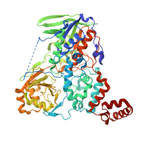 | |
UniProt | |||||
Entity Groups | |||||
| Sequence Clusters | 30% Identity50% Identity70% Identity90% Identity95% Identity100% Identity | ||||
| UniProt Group | Q2G1P3 | ||||
Sequence AnnotationsExpand | |||||
Reference Sequence | |||||
| Ligands 3 Unique | |||||
|---|---|---|---|---|---|
| ID | Chains | Name / Formula / InChI Key | 2D Diagram | 3D Interactions | |
| EIC (Subject of Investigation/LOI) Download:Ideal Coordinates CCD File | D [auth A], F [auth B], I [auth C] | LINOLEIC ACID C18 H32 O2 OYHQOLUKZRVURQ-HZJYTTRNSA-N |  | ||
| PEG Download:Ideal Coordinates CCD File | E [auth A], G [auth B], H [auth B], J [auth C] | DI(HYDROXYETHYL)ETHER C4 H10 O3 MTHSVFCYNBDYFN-UHFFFAOYSA-N |  | ||
| GOL Download:Ideal Coordinates CCD File | K [auth C] | GLYCEROL C3 H8 O3 PEDCQBHIVMGVHV-UHFFFAOYSA-N |  | ||
| Length ( Å ) | Angle ( ˚ ) |
|---|---|
| a = 189.355 | α = 90 |
| b = 114.114 | β = 117.04 |
| c = 119.113 | γ = 90 |
| Software Name | Purpose |
|---|---|
| PHENIX | refinement |
| HKL-3000 | data reduction |
| HKL-3000 | data scaling |
| PHENIX | phasing |
| Funding Organization | Location | Grant Number |
|---|---|---|
| Other government | China | 2021YFC2103702 |


