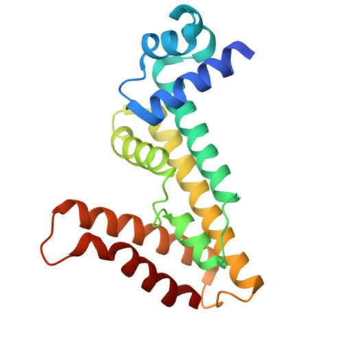
Structural basis for spermidine recognition and modulation of Acinetobacter baumannii multidrug efflux regulator AmvR.
Wang, N., Wang, X., Zhou, M., Lu, Q., Xu, Y., Wang, Y., Wang, H., Yang, B., He, S., Xu, L., Li, J., Ge, H., Ma, J.(2025) mBio 16: e0008125-e0008125
- PubMed: 40162807 Search on PubMedSearch on PubMed Central
- DOI: https://doi.org/10.1128/mbio.00081-25
- Primary Citation Related Structures:
8WP6, 9IPT - PubMed Abstract:
Acinetobacter baumannii is a gram-negative, opportunistic pathogen frequently associated with hospital-acquired infections. Due to its resistance to multiple antibiotics, it is emerging as a major nosocomial pathogen, causing a wide range of severe infections such as pneumonia, meningitis, and bloodstream infections. In many cases, the intrinsic activities of efflux pumps contribute to the development of drug resistance. The polyamine-binding protein AmvR regulates the multidrug efflux pump AmvA, which is pivotal for transporting polyamines, an abundant and prevalent class of amino acid-derived metabolites. Here, we report the crystal structure of the AmvR protein bound to its physiological substrate, spermidine, thereby offering structural and functional insights into AmvR. By employing electrophoretic mobility shift assays and DNase I footprinting, we identified the recognition sites of the intragenic regions of amvR and amvA by AmvR. Moreover, a fluorescence reporter assay revealed that AmvR repressed the expressions of AmvA and AmvR. In addition, isothermal titration calorimetry indicated that spermidine may be a natural ligand of AmvR. Collectively, these experiments provided a better understanding of substrate recognition for the discovery of potential inhibitors. Furthermore, our results revealed that substrate binding triggers a localized conformational change in the AmvR protein, as supported by size-exclusion chromatography and static light scattering, suggesting a distinctive regulatory mechanism within the TetR family transcription factors. Multidrug efflux pumps are key contributors to clinically significant drug resistance in various gram-negative pathogens responsible for hospital-acquired infections. These pathogens often possess multiple genes that encode potential multidrug efflux pumps. Identifying the specific regulatory proteins that control the expression of these pumps, along with elucidating the regulatory mechanisms triggered by effectors, presents a complex challenge. In this study, we have resolved the crystal structures of AmvR in both its unbound and spermidine-bound states. To the best of our knowledge, this represents the first validated structural model of a polyamine-bound transcriptional regulator. Through detailed structural analysis and functional assays, we have pinpointed the critical residues in AmvR responsible for substrate recognition, providing a foundation for the development of future inhibitors.
- Institute of Health Sciences and Technology, Institutes of Physical and Information Technology, Anhui University, Hefei, Anhui, China.
Organizational Affiliation: