9IP8 | pdb_00009ip8

Poly-alanine model for HL-type bispecific diabody Ex3 composed of 528 and OKT3 Fvs in ternary complex with sEGFR and CD3gamma-epsilon (closed conformation)


Experimental Data Snapshot

  • Method: ELECTRON MICROSCOPY
  • Resolution: 3.91 Å
  • Aggregation State: PARTICLE 
  • Reconstruction Method: SINGLE PARTICLE 

wwPDB Validation 3D Report Full Report

Validation slider image for 9IP8

This is version 1.3 of the entry. See complete history. 

Literature

Bispecific antibody-antigen complex structures reveal activity enhancement by domain rearrangement.

Sato, K., Uehara, S., Tsugita, A., Ishii, M., Ishiyama, S., Maejima, A., Nakahara, I., Nazuka, M., Matsui, T., Christos, G., Yokoyama, T., Kumagai, I., Makabe, K., Asano, R., Tanaka, Y.

(2025) Cell Rep 44: 115965-115965

  • DOI: https://doi.org/10.1016/j.celrep.2025.115965
  • Primary Citation Related Structures: 
    9IP7, 9IP8, 9IP9, 9IPA, 9IPB, 9IPC, 9IPD, 9IPE

  • PubMed Abstract: 

    Bispecific antibodies (BsAbs) have been developed as anti-cancer drugs that accumulate activated T cells on cancer cells by bridging the antigens present in each cell. Ex3 is a diabody-type BsAb composed of an anti-epidermal growth factor receptor (EGFR) antibody and an anti-CD3 antibody. In the design of Ex3, the LH-type domain order (Ex3LH) is shown to have more than 100-fold greater anti-cancer activity than the HL-type domain order (Ex3HL). To understand this phenomenon of activity enhancement by domain-order rearrangement, we report here cryoelectron microscopy (cryo-EM) structures of both Ex3HL and Ex3LH in complex with EGFR and CD3. A structural comparison of the HL and LH types reveals that the domain rearrangement leads to drastic structural changes and that the avoidance of steric hindrance by a favorable bridging angle on the cell surface is the fundamental mechanism for this activity enhancement.


  • Organizational Affiliation: 
    • Graduate School of Life Sciences, Tohoku University, Miyagi 980-8577, Japan.

Macromolecule Content 

  • Total Structure Weight: 149.22 kDa 
  • Atom Count: 5,610 
  • Modeled Residue Count: 1,143 
  • Deposited Residue Count: 1,358 
  • Unique protein chains: 3

Macromolecules

Find similar proteins by:|  3D Structure
Entity ID: 1
MoleculeChains  Sequence LengthOrganismDetailsImage
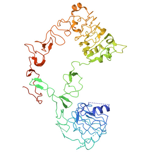
Epidermal growth factor receptor627Homo sapiensMutation(s): 0 
Gene Names: EGFR, ERBB, ERBB1, HER1
EC: 2.7.10.1
UniProt & NIH Common Fund Data Resources
Find proteins for P00533 (Homo sapiens)
Explore P00533 
Go to UniProtKB:  P00533
PHAROS:  P00533
GTEx:  ENSG00000146648 
Entity Groups
Sequence Clusters30% Identity50% Identity70% Identity90% Identity95% Identity100% Identity
UniProt GroupP00533
Sequence Annotations
Expand
Reference Sequence
Find similar proteins by:|  3D Structure
Entity ID: 2
MoleculeChains  Sequence LengthOrganismDetailsImage
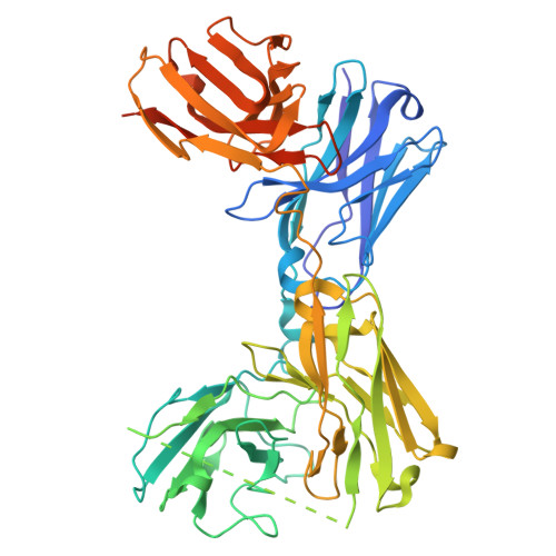
HL-type bispecific diabody Ex3527synthetic constructMutation(s): 0 
Entity Groups
Sequence Clusters30% Identity50% Identity70% Identity90% Identity95% Identity100% Identity
Sequence Annotations
Expand
Reference Sequence
Find similar proteins by:|  3D Structure
Entity ID: 3
MoleculeChains  Sequence LengthOrganismDetailsImage
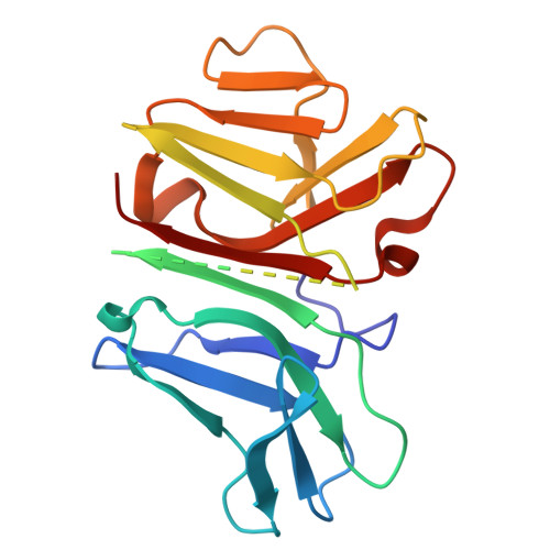
T-cell surface glycoprotein CD3 gamma chain,T-cell surface glycoprotein CD3 epsilon chain204Homo sapiensMutation(s): 0 
Gene Names: CD3G, T3G, CD3E, T3E
UniProt & NIH Common Fund Data Resources
Find proteins for P07766 (Homo sapiens)
Explore P07766 
Go to UniProtKB:  P07766
PHAROS:  P07766
GTEx:  ENSG00000198851 
Find proteins for P09693 (Homo sapiens)
Explore P09693 
Go to UniProtKB:  P09693
PHAROS:  P09693
GTEx:  ENSG00000160654 
Entity Groups
Sequence Clusters30% Identity50% Identity70% Identity90% Identity95% Identity100% Identity
UniProt GroupsP07766P09693
Sequence Annotations
Expand
Reference Sequence

Experimental Data & Validation

Experimental Data

  • Method: ELECTRON MICROSCOPY
  • Resolution: 3.91 Å
  • Aggregation State: PARTICLE 
  • Reconstruction Method: SINGLE PARTICLE 
EM Software:
TaskSoftware PackageVersion
MODEL REFINEMENTPHENIX

Structure Validation

View Full Validation Report


Entry History 

& Funding Information

Deposition Data


Funding OrganizationLocationGrant Number
Japan Society for the Promotion of Science (JSPS)Japan--

Revision History  (Full details and data files)

  • Version 1.0: 2025-05-28
    Type: Initial release
  • Version 1.1: 2025-07-02
    Changes: Data collection
  • Version 1.2: 2025-07-30
    Changes: Data collection, Database references
  • Version 1.3: 2025-08-06
    Changes: Data collection, Database references