9INC | pdb_00009inc

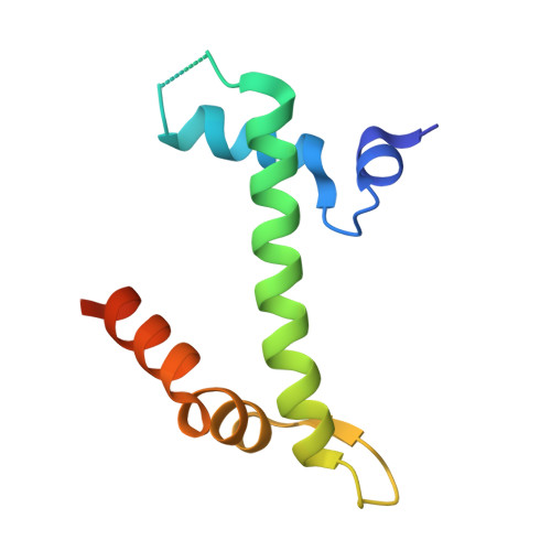
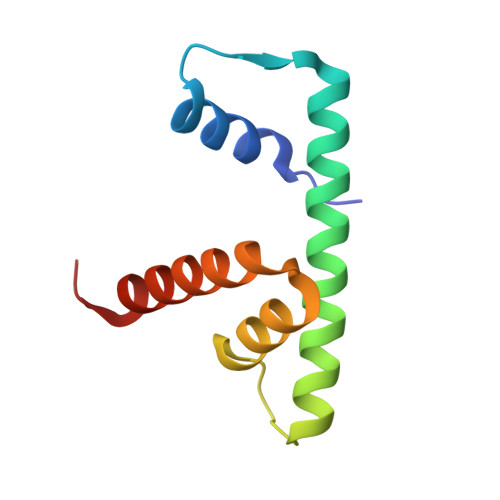
High resolutional Crystal Structure of human H2A.Z-H2B dimer in complex with human YL1-Z domain


Experimental Data Snapshot

  • Method: X-RAY DIFFRACTION
  • Resolution: 2.01 Å
  • R-Value Free: 
    0.257 (Depositor), 0.256 (DCC) 
  • R-Value Work: 
    0.220 (Depositor), 0.220 (DCC) 
  • R-Value Observed: 
    0.221 (Depositor) 

Starting Models: experimental
View more details

wwPDB Validation 3D Report Full Report

Validation slider image for 9INC

This is version 1.1 of the entry. See complete history

Literature

Structural insights into specific recognition of the histone variant H2A.Z by the YL1 subunit of SRCAP chromatin-remodeling complex.

Tan, W.Liu, Y.Hong, J.

(2026) J Biol Chem 302: 111294-111294

  • DOI: https://doi.org/10.1016/j.jbc.2026.111294
  • Primary Citation Related Structures: 
    9INC

  • PubMed Abstract: 

    The SNF2-related CREB-binding protein activator protein complex regulates gene transcription through chromatin remodeling, where the subunit YL1 mediates the exchange of H2A-H2B dimers with H2A.Z-H2B in the nucleosome. Although structures of YL1 in complex with H2A.Z-H2B have been previously reported for both Drosophila and Homo sapiens, the complex structures of the two species exhibit significant differences, which have caused some confusion over how YL1 recruits H2A.Z-H2B. We determined the high-resolution (2.01 Å) crystal structure of the N-terminal region (8-73) of the human SNF2-related CREB-binding protein activator protein subunit YL1 in complex with H2B-H2A.Z; this domain is termed as YL1-Z. YL1-Z consists of an alpha helix and a longer loop, which bind to H2A.Z-H2B to form a complex. In the structure, residues Phe29, Tyr30, Tyr34, and Phe37 of the YL1-Z domain specifically recognize the hydrophobic residues, Gln87, Ile90, Ile100, and Ile104, of H2A.Z through hydrophobic interactions, forming a hydrophobic core. In addition, residues Asp55, Asp58, and Asp60 of YL1-Z form electrostatic interactions with Arg34 and Lys37 on H2A.Z, stabilizing the complex structure. The particular recognition has been validated in vitro by maltose-binding protein-pulldown and isothermal titration calorimetry experiments. Our data show that YL1 employs a conserved structural mechanism for recognizing the H2A.Z-H2B dimer.


  • Organizational Affiliation
    • Institute of Health Sciences and Technology (IHST), Institutes of Physical Sciences and Information Technology, Anhui University, Hefei, China.

Macromolecule Content 

  • Total Structure Weight: 61.02 kDa 
  • Atom Count: 3,735 
  • Modeled Residue Count: 475 
  • Deposited Residue Count: 558 
  • Unique protein chains: 3

Macromolecules

Find similar proteins by:|  3D Structure
Entity ID: 1
MoleculeChains  Sequence LengthOrganismDetailsImage
Histone H2A.Z
A, D
98Homo sapiensMutation(s): 0 
Gene Names: H2AZ1H2AFZH2AZ
UniProt & NIH Common Fund Data Resources
Find proteins for P0C0S5 (Homo sapiens)
Explore P0C0S5 
Go to UniProtKB:  P0C0S5
PHAROS:  P0C0S5
GTEx:  ENSG00000164032 
Entity Groups
Sequence Clusters30% Identity50% Identity70% Identity90% Identity95% Identity100% Identity
UniProt GroupP0C0S5
Sequence Annotations
Expand
Reference Sequence
Find similar proteins by:|  3D Structure
Entity ID: 2
MoleculeChains  Sequence LengthOrganismDetailsImage
Histone H2B type 1-J
B, E
94Homo sapiensMutation(s): 0 
Gene Names: H2BC11H2BFRHIST1H2BJ
UniProt & NIH Common Fund Data Resources
Find proteins for P06899 (Homo sapiens)
Explore P06899 
Go to UniProtKB:  P06899
PHAROS:  P06899
GTEx:  ENSG00000124635 
Entity Groups
Sequence Clusters30% Identity50% Identity70% Identity90% Identity95% Identity100% Identity
UniProt GroupP06899
Sequence Annotations
Expand
Reference Sequence
Find similar proteins by:|  3D Structure
Entity ID: 3
MoleculeChains  Sequence LengthOrganismDetailsImage
Vacuolar protein sorting-associated protein 72 homolog
C, F
87Homo sapiensMutation(s): 0 
Gene Names: VPS72TCFL1YL1
UniProt & NIH Common Fund Data Resources
Find proteins for Q15906 (Homo sapiens)
Explore Q15906 
Go to UniProtKB:  Q15906
PHAROS:  Q15906
GTEx:  ENSG00000163159 
Entity Groups
Sequence Clusters30% Identity50% Identity70% Identity90% Identity95% Identity100% Identity
UniProt GroupQ15906
Sequence Annotations
Expand
Reference Sequence

Experimental Data & Validation

Experimental Data

  • Method: X-RAY DIFFRACTION
  • Resolution: 2.01 Å
  • R-Value Free:  0.257 (Depositor), 0.256 (DCC) 
  • R-Value Work:  0.220 (Depositor), 0.220 (DCC) 
  • R-Value Observed: 0.221 (Depositor) 
Space Group: C 2 2 21
Unit Cell:
Length ( Å )Angle ( ˚ )
a = 116.093α = 90
b = 126.99β = 90
c = 70.757γ = 90
Software Package:
Software NamePurpose
PHENIXrefinement
PDB_EXTRACTdata extraction
DIALSdata reduction
DIALSdata scaling
PHENIXphasing

Structure Validation

View Full Validation Report


Entry History 

& Funding Information

Deposition Data


Funding OrganizationLocationGrant Number
National Natural Science Foundation of China (NSFC)China31970669

Revision History  (Full details and data files)

  • Version 1.0: 2025-07-09
    Type: Initial release
  • Version 1.1: 2026-07-22
    Changes: Database references