


Crystal structure of the periplasmic domain of sensor protein EvgS from Escherichia coli str. K-12 substr. MG1655
Teng, Y., Liu, R., Luo, B.To be published.
Experimental Data Snapshot
Starting Model: in silico
View more details
wwPDB Validation 3D Report Full Report

Entity ID: 1 | |||||
|---|---|---|---|---|---|
| Molecule | Chains | Sequence Length | Organism | Details | Image |
| Acid-sensing system histidine kinase EvgS | 513 | Escherichia coli | Mutation(s): 0 Gene Names: evgS, GP975_24990 EC: 2.7.13.3 | 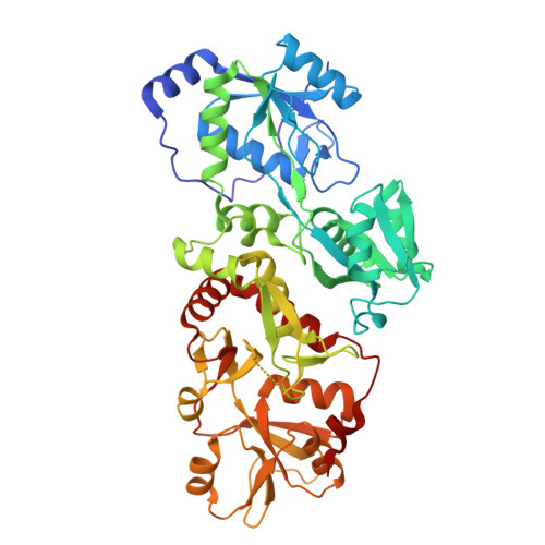 | |
UniProt | |||||
Entity Groups | |||||
| Sequence Clusters | 30% Identity50% Identity70% Identity90% Identity95% Identity100% Identity | ||||
| UniProt Group | P30855 | ||||
Sequence AnnotationsExpand | |||||
Reference Sequence | |||||
| Length ( Å ) | Angle ( ˚ ) |
|---|---|
| a = 85.56 | α = 90 |
| b = 109.914 | β = 90 |
| c = 118.666 | γ = 90 |
| Software Name | Purpose |
|---|---|
| PHENIX | refinement |
| XDS | data scaling |
| XDS | data reduction |
| PHASER | phasing |
| Funding Organization | Location | Grant Number |
|---|---|---|
| Not funded | -- |


