9IJO | pdb_00009ijo

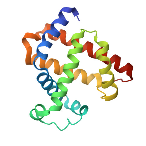
X-ray crystal structure of F46C myoglobin with a covalently linked 4-methyl-2,2'-bipyridine group in complex with Cu2+


Experimental Data Snapshot

  • Method: X-RAY DIFFRACTION
  • Resolution: 1.53 Å
  • R-Value Free: 
    0.194 (Depositor), 0.194 (DCC) 
  • R-Value Work: 
    0.167 (Depositor), 0.168 (DCC) 
  • R-Value Observed: 
    0.169 (Depositor) 

Starting Model: experimental
View more details

wwPDB Validation 3D Report Full Report

Validation slider image for 9IJO

Ligand Structure Quality Assessment 


This is version 1.1 of the entry. See complete history

Literature

Rational Design of an Artificial Metalloenzyme by Constructing a Metal-Binding Site Close to the Heme Cofactor in Myoglobin.

Nie, L.S.Liu, X.C.Yu, L.Liu, A.K.Sun, L.J.Gao, S.Q.Lin, Y.W.

(2024) Inorg Chem 63: 18531-18535

  • DOI: https://doi.org/10.1021/acs.inorgchem.4c03093
  • Primary Citation Related Structures: 
    9IJO

  • PubMed Abstract: 

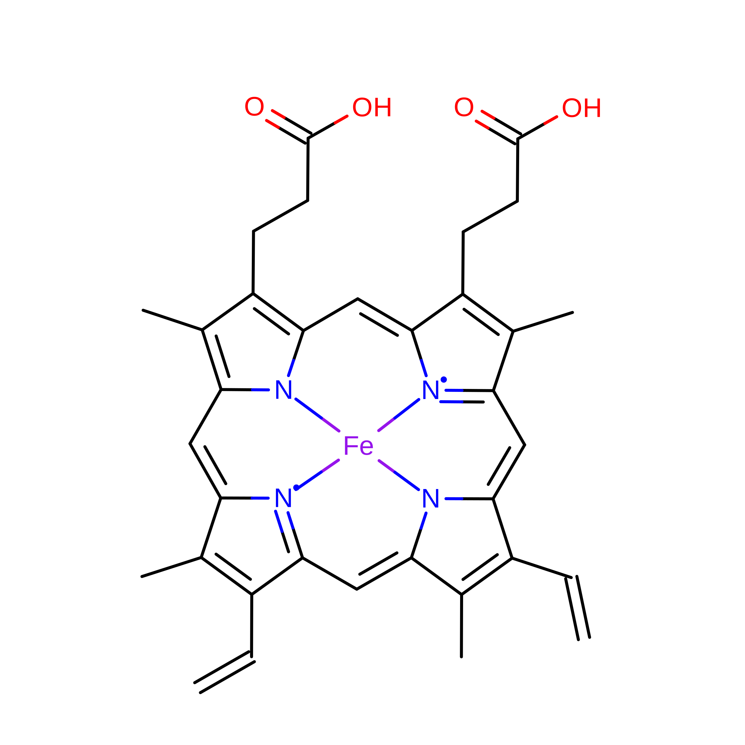
    In this study, we constructed a metal-binding site close to the heme cofactor in myoglobin (Mb) by covalently attaching a nonnative metal-binding ligand of bipyridine to Cys46 through the F46C mutation in the heme distal site. The X-ray structure of the designed enzyme, termed F46C-mBpy Mb, was solved in the Cu(II)-bound form, which revealed the formation of a heterodinuclear center of Cu-His-H 2 O-heme. Cu(II)-F46C-mBpy Mb exhibits not only nitrite reductase reactivity but also cascade reaction activity involving both hydrolysis and oxidation. Furthermore, F46C-mBpy Mb displays Mn-peroxidase activity by the oxidation of Mn 2+ to Mn 3+ using H 2 O 2 as an oxidant. This study shows that the construction of a nonnative metal-binding site close to the heme cofactor is a convenient approach to creating an artificial metalloenzyme with a heterodinuclear center that confers multiple functions.


  • Organizational Affiliation
    • School of Chemistry and Chemical Engineering, University of South China, Hengyang 421001, China.

Macromolecule Content 

  • Total Structure Weight: 18.19 kDa 
  • Atom Count: 1,463 
  • Modeled Residue Count: 152 
  • Deposited Residue Count: 154 
  • Unique protein chains: 1

Macromolecules

Find similar proteins by:|  3D Structure
Entity ID: 1
MoleculeChains  Sequence LengthOrganismDetailsImage
Myoglobin154Physeter macrocephalusMutation(s): 1 
Gene Names: MB
EC: 1.11.1 (UniProt), 1.7 (UniProt)
UniProt
Find proteins for P02185 (Physeter macrocephalus)
Explore P02185 
Go to UniProtKB:  P02185
Entity Groups
Sequence Clusters30% Identity50% Identity70% Identity90% Identity95% Identity100% Identity
UniProt GroupP02185
Sequence Annotations
Expand
Reference Sequence

Experimental Data & Validation

Experimental Data

  • Method: X-RAY DIFFRACTION
  • Resolution: 1.53 Å
  • R-Value Free:  0.194 (Depositor), 0.194 (DCC) 
  • R-Value Work:  0.167 (Depositor), 0.168 (DCC) 
  • R-Value Observed: 0.169 (Depositor) 
Space Group: P 21 21 21
Unit Cell:
Length ( Å )Angle ( ˚ )
a = 48.989α = 90
b = 77.523β = 90
c = 39.751γ = 90
Software Package:
Software NamePurpose
PHENIXrefinement
PHENIXrefinement
xia2data reduction
DIALSdata scaling
PHASERphasing

Structure Validation

View Full Validation Report


Ligand Structure Quality Assessment 


Entry History 

& Funding Information

Deposition Data

  • Released Date: 2025-07-02 
  • Deposition Author(s): Lin, Y.W.

Funding OrganizationLocationGrant Number
National Science Foundation (NSF, China)China--

Revision History  (Full details and data files)

  • Version 1.0: 2025-07-02
    Type: Initial release
  • Version 1.1: 2026-07-15
    Changes: Database references