9IJB | pdb_00009ijb

Crystal structure and function analysis of a highly potential drug target 6-phosphogluconate dehydrogenase in Mycobacterium tuberculosis


Experimental Data Snapshot

  • Method: X-RAY DIFFRACTION
  • Resolution: 2.74 Å
  • R-Value Free: 
    0.251 (Depositor), 0.250 (DCC) 
  • R-Value Work: 
    0.198 (Depositor), 0.200 (DCC) 
  • R-Value Observed: 
    0.201 (Depositor) 

Starting Model: experimental
View more details

wwPDB Validation   3D Report Full Report


This is version 1.1 of the entry. See complete history


Literature

Crystal structure and function analysis of 6-phosphogluconate dehydrogenase in Mycobacterium tuberculosis.

Wang, Y.Ren, X.Li, T.Su, D.Zhang, R.

(2024) Biochem Biophys Res Commun 731: 150390-150390

  • DOI: https://doi.org/10.1016/j.bbrc.2024.150390
  • Primary Citation of Related Structures:  
    9IJB

  • PubMed Abstract: 

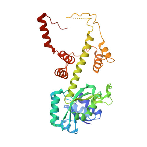
    6-phosphogluconate dehydrogenase (6PGDH) is an essential enzyme in energy metabolism and redox reactions, and represents a potential drug target for the development of therapies targeting trypanosomes, plasmodium, or other pathogens. Tuberculosis, caused by Mycobacterium tuberculosis, is a contagious disease that severely affects human health, with approximately one-third of the world's population infected. However, the protein structure, exact oligomeric state, and catalytic mechanism of 6PGDH in Mycobacterium tuberculosis (Mt6PGDH) have remained largely unknown. In this study, we successfully purified and determined the structure of Mt6PGDH, revealing its function as a tetramer in both solution and crystal states. Through structural comparisons, we clarified the tetramer formation mechanism and the oligomeric organization of short-chain 6PGDHs. Additionally, we identified key residues for coenzyme recognition and catalytic activity. This work not only deepens our understanding of the enzymatic function of Mt6PGDH but also lays a foundation for the development of drugs targeting this enzyme.


  • Organizational Affiliation
    • Department of Ophthalmology, West China Hospital, Sichuan University, Chengdu, 610041, PR China; State Key Laboratory of Biotherapy and Collaborative Innovation Center for Biotherapy, West China Hospital, Sichuan University, Chengdu, 610041, PR China.

Macromolecules
Find similar proteins by:  (by identity cutoff)  |  3D Structure
Entity ID: 1
MoleculeChains Sequence LengthOrganismDetailsImage
6-phosphogluconate dehydrogenase, NAD(+)-dependent, decarboxylating
A, B, C, D
340Mycobacterium tuberculosisMutation(s): 0 
Gene Names: 
EC: 1.1.1.343 (PDB Primary Data), 1.1.1.44 (PDB Primary Data)
UniProt
Find proteins for A0A045IPA0 (Mycobacterium tuberculosis)
Explore A0A045IPA0 
Go to UniProtKB:  A0A045IPA0
Entity Groups  
Sequence Clusters30% Identity50% Identity70% Identity90% Identity95% Identity100% Identity
UniProt GroupA0A045IPA0
Sequence Annotations
Expand
  • Reference Sequence
Experimental Data & Validation

Experimental Data

  • Method: X-RAY DIFFRACTION
  • Resolution: 2.74 Å
  • R-Value Free:  0.251 (Depositor), 0.250 (DCC) 
  • R-Value Work:  0.198 (Depositor), 0.200 (DCC) 
  • R-Value Observed: 0.201 (Depositor) 
Space Group: P 21 21 21
Unit Cell:
Length ( Å )Angle ( ˚ )
a = 95.991α = 90
b = 107.246β = 90
c = 137.023γ = 90
Software Package:
Software NamePurpose
PHENIXrefinement
HKL-2000data scaling
HKL-2000data reduction
PHASERphasing

Structure Validation

View Full Validation Report


Entry History & Funding Information

Deposition Data


Funding OrganizationLocationGrant Number
National Natural Science Foundation of China (NSFC)China2017YFA0504300
National Natural Science Foundation of China (NSFC)China2017YFA0505900
National Natural Science Foundation of China (NSFC)China32360046

Revision History  (Full details and data files)

  • Version 1.0: 2024-07-03
    Type: Initial release
  • Version 1.1: 2024-08-07
    Changes: Database references