STK4 inhibits the E3 activity of HOIP by phosphorylating its allosteric ubiquitin-binding site.
Wang, Y., Zhou, X., Lin, Z., Huang, Y., Zhang, Y., Liu, H., Zhou, Y., Liu, J., Pan, L.(2025) Cell Discov 11: 75-75
- PubMed: 40957900 Search on PubMedSearch on PubMed Central
- DOI: https://doi.org/10.1038/s41421-025-00824-x
- Primary Citation Related Structures:
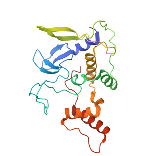
9IIC - PubMed Abstract:
HOIP, an RBR-type E3 ligase and the catalytic subunit of the linear ubiquitin chain assembly complex (LUBAC), plays crucial roles in various cellular processes, including the NF-κB signaling pathway. The E3 activity of HOIP can be inhibited by the kinase STK4-mediated phosphorylation, although the mechanism is poorly understood. In this study, using biochemical, mass spectrometry and structural approaches, we systemically characterize the association of STK4 with HOIP, and unveil that STK4 can directly bind to the RING2-LDD module of HOIP through its kinase domain. The determined crystal structure of STK4 in complex with HOIP RING2-LDD not only elucidates the detailed binding mechanism of STK4 with HOIP, but also uncovers, for the first time, a unique binding mode of STK4 with its substrate. Moreover, we reveal that STK4 can directly phosphorylate the T786 residue of HOIP that is located in the allosteric ubiquitin-binding site of HOIP. Importantly, the phosphorylation of HOIP T786 mediated by STK4 can block the binding of ubiquitin to the allosteric site of HOIP, thereby attenuating the E3 activity of HOIP. In all, our findings provide mechanistic insights into the interaction between STK4 and HOIP as well as the negative regulation of HOIP's E3 activity by STK4-mediated phosphorylation, which are valuable for further understanding the regulatory modes of RBR-type E3 ligases.
- School of Chemistry and Materials Science, Hangzhou Institute for Advanced Study, University of Chinese Academy of Sciences, 1 Sub-lane Xiangshan, Hangzhou, Zhejiang, China.
Organizational Affiliation: