9ICR

DNA POLYMERASE BETA (E.C.2.7.7.7)/DNA COMPLEX + 2'-DEOXYCYTIDINE-5'-TRIPHOSPHATE, SOAKED IN THE PRESENCE OF DCTP AND MNCL2


Experimental Data Snapshot

  • Method: X-RAY DIFFRACTION
  • Resolution: 3.00 Å
  • R-Value Observed: 0.158 

wwPDB Validation   3D Report Full Report


Ligand Structure Quality Assessment 


This is version 1.2 of the entry. See complete history


Literature

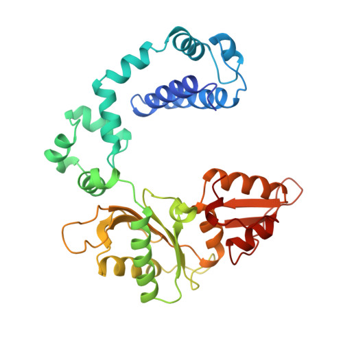
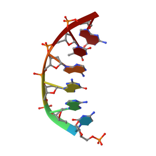
A structural basis for metal ion mutagenicity and nucleotide selectivity in human DNA polymerase beta

Pelletier, H.Sawaya, M.R.Wolfle, W.Wilson, S.H.Kraut, J.

(1996) Biochemistry 35: 12762-12777

  • DOI: https://doi.org/10.1021/bi9529566
  • Primary Citation of Related Structures:  
    1ZQT, 7ICE, 7ICF, 7ICG, 7ICH, 7ICI, 7ICJ, 7ICK, 7ICL, 7ICM, 7ICN, 7ICO, 7ICP, 7ICQ, 7ICR, 7ICS, 7ICT, 7ICU, 7ICV, 8ICJ, 8ICK, 8ICL, 8ICM, 8ICN, 8ICO, 8ICP, 8ICQ, 8ICR, 8ICS, 8ICT, 8ICU, 8ICV, 8ICW, 8ICX, 8ICY, 9ICF, 9ICK, 9ICN, 9ICO, 9ICP, 9ICQ, 9ICR, 9ICS, 9ICT, 9ICU, 9ICV

  • PubMed Abstract: 
  • When crystals of human DNA polymerase beta (pol beta) complexed with DNA [Pelletier, H., Sawaya, M. R., Wolfle, W., Wilson, S. H., & Kraut, J. (1996) Biochemistry 35, 12742-12761] are soaked in the presence of dATP and Mn2+, X-ray structural analysis shows that nucleotidyl transfer to the primer 3'-OH takes place directly in the crystals, even though the DNA is blunt-ended at the active site ...

    When crystals of human DNA polymerase beta (pol beta) complexed with DNA [Pelletier, H., Sawaya, M. R., Wolfle, W., Wilson, S. H., & Kraut, J. (1996) Biochemistry 35, 12742-12761] are soaked in the presence of dATP and Mn2+, X-ray structural analysis shows that nucleotidyl transfer to the primer 3'-OH takes place directly in the crystals, even though the DNA is blunt-ended at the active site. Under similar crystal-soaking conditions, there is no evidence for a reaction when Mn2+ is replaced by Mg2+, which is thought to be the divalent metal ion utilized by most polymerases in vivo. These results suggest that one way Mn2+ may manifest its mutagenic effect on polymerases is by promoting greater reactivity than Mg2+ at the catalytic site, thereby allowing the nucleotidyl transfer reaction to take place with little or no regard to instructions from a template. Non-template-directed nucleotidyl transfer is also observed when pol beta-DNA cocrystals are soaked in the presence of dATP and Zn2+, but the reaction products differ in that the sugar moiety of the incorporated nucleotide appears distorted or otherwise cleaved, in agreement with reports that Zn2+ may act as a polymerase inhibitor rather than as a mutagen [Sirover, M. A., & Loeb, L. A. (1976) Science 194, 1434-1436]. Although no reaction is observed when crystals are soaked in the presence of dATP and other metal ions such as Ca2+, Co2+, Cr3+, or Ni2+, X-ray structural analyses show that these metal ions coordinate the triphosphate moiety of the nucleotide in a manner that differs from that observed with Mg2+. In addition, all metal ions tested, with the exception of Mg2+, promote a change in the side-chain position of aspartic acid 192, which is one of three highly conserved active-site carboxylate residues. Soaking experiments with nucleotides other than dATP (namely, dCTP, dGTP, dTTP, ATP, ddATP, ddCTP, AZT-TP, and dATP alpha S) reveal a non-base-specific binding site on pol beta for the triphosphate and sugar moieties of a nucleotide, suggesting a possible mechanism for nucleotide selectivity whereby triphosphate-sugar binding precedes a check for correct base pairing with the template.


    Related Citations: 
    • Crystal Structures of Human DNA Polymerase Beta Complexed with Nicked and Gapped DNA Substrates
      Sawaya, M.R., Rawson, T., Wilson, S.H., Kraut, J., Pelletier, H.
      () To be published --: --
    • The Role of Thumb Movement and Template Bending in Polymerase Fidelity
      Pelletier, H.
      () To be published --: --
    • Crystal Structures of Human DNA Polymerase Beta Complexed with DNA; Implications for Catalytic Mechanism, Processivity, and Fidelity
      Pelletier, H., Sawaya, M.R., Wolfle, W., Wilson, S.H., Kraut, J.
      (1996) Biochemistry 35: 12742
    • Characterization of the Metal Ion-Binding HHH Motifs in Human DNA Polymerase Beta by X-Ray Structural Analysis
      Pelletier, H., Sawaya, M.R.
      (1996) Biochemistry 35: 12778
    • Polymerase Structures and Mechanism
      Pelletier, H.
      (1994) Science 266: 2025
    • Structures of Ternary Complexes of Rat DNA Polymerase Beta, a DNA Template- Primer, and ddCTP
      Pelletier, H., Sawaya, M.R., Kumar, A., Wilson, S.H., Kraut, J.
      (1994) Science 264: 1891
    • Crystal Structure of Rat DNA Polymerase Beta: Evidence for a Common Polymerase Mechanism
      Sawaya, M.R., Pelletier, H., Kumar, A., Wilson, S.H., Kraut, J.
      (1994) Science 264: 1930

    Organizational Affiliation

    Department of Chemistry and Biochemistry, University of California, San Diego, La Jolla 92093-0506, USA.


Macromolecules

Find similar proteins by:  (by identity cutoff)  |  3D Structure
Entity ID: 3
MoleculeChainsSequence LengthOrganismDetailsImage
PROTEIN (DNA POLYMERASE BETA (E.C.2.7.7.7))C [auth A]335Homo sapiensMutation(s): 0 
Gene Names: POLB
EC: 2.7.7.7 (UniProt), 4.2.99 (UniProt)
UniProt & NIH Common Fund Data Resources
Find proteins for P06746 (Homo sapiens)
Explore P06746 
Go to UniProtKB:  P06746
PHAROS:  P06746
Entity Groups  
Sequence Clusters30% Identity50% Identity70% Identity90% Identity95% Identity100% Identity
UniProt GroupP06746
Protein Feature View
Expand
  • Reference Sequence

Find similar nucleic acids by:  Sequence   |   3D Structure  

Entity ID: 1
MoleculeChainsLengthOrganismImage
DNA (5'-D(*CP*AP*TP*CP*TP*GP*T)-3')A [auth T]7N/A
Protein Feature View
Expand
  • Reference Sequence

Find similar nucleic acids by:  Sequence   |   3D Structure  

Entity ID: 2
MoleculeChainsLengthOrganismImage
DNA (5'-D(*CP*AP*GP*AP*TP*G)-3')B [auth P]6N/A
Protein Feature View
Expand
  • Reference Sequence
Small Molecules
Ligands 3 Unique
IDChainsName / Formula / InChI Key2D Diagram3D Interactions
DCP
Query on DCP

Download Ideal Coordinates CCD File 
H [auth A]2'-DEOXYCYTIDINE-5'-TRIPHOSPHATE
C9 H16 N3 O13 P3
RGWHQCVHVJXOKC-SHYZEUOFSA-N
 Ligand Interaction
MN
Query on MN

Download Ideal Coordinates CCD File 
D [auth A],
E [auth A]
MANGANESE (II) ION
Mn
WAEMQWOKJMHJLA-UHFFFAOYSA-N
 Ligand Interaction
NA
Query on NA

Download Ideal Coordinates CCD File 
F [auth A],
G [auth A]
SODIUM ION
Na
FKNQFGJONOIPTF-UHFFFAOYSA-N
 Ligand Interaction
Experimental Data & Validation

Experimental Data

  • Method: X-RAY DIFFRACTION
  • Resolution: 3.00 Å
  • R-Value Observed: 0.158 
  • Space Group: P 21 21 2
Unit Cell:
Length ( Å )Angle ( ˚ )
a = 178.602α = 90
b = 57.637β = 90
c = 48.459γ = 90
Software Package:
Software NamePurpose
TNTrefinement
SDMSdata reduction
SDMSdata scaling
TNTphasing

Structure Validation

View Full Validation Report


Ligand Structure Quality Assessment 


Entry History 

Deposition Data

Revision History  (Full details and data files)

  • Version 1.0: 1996-01-04
    Type: Initial release
  • Version 1.1: 2008-05-22
    Changes: Version format compliance
  • Version 1.2: 2011-07-13
    Changes: Version format compliance