9IC4 | pdb_00009ic4

Crystal structure of beta-lactamase-like domain of ComEC from Moorella glycerini


Experimental Data Snapshot

  • Method: X-RAY DIFFRACTION
  • Resolution: 1.86 Å
  • R-Value Free: 
    0.203 (Depositor), 0.205 (DCC) 
  • R-Value Work: 
    0.170 (Depositor), 0.172 (DCC) 
  • R-Value Observed: 
    0.171 (Depositor) 

Starting Model: in silico
View more details

wwPDB Validation   3D Report Full Report


This is version 1.1 of the entry. See complete history


Literature

Molecular interplay between ComEC domains allows for selective degradation of the non-translocating strand during natural transformation.

Stedman, M.J.M.Deselaers, S.Braus, S.A.G.Wang, D.Balaguer, M.G.Gossert, A.D.Hospenthal, M.K.

(2025) Nucleic Acids Res 53

  • DOI: https://doi.org/10.1093/nar/gkaf932
  • Primary Citation of Related Structures:  
    9IC4, 9IEW

  • PubMed Abstract: 

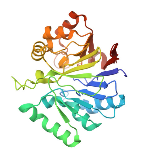
    Naturally competent bacteria can take up and incorporate environmental DNA using complex machinery in a process called natural transformation. This is a key mechanism in the spread of antibiotic resistance amongst bacteria, including many human pathogens. All competent bacteria require ComEC to transport the transforming DNA across the cytoplasmic membrane. In addition to the transmembrane domain predicted to form the DNA channel, most ComEC orthologues contain an oligonucleotide binding (OB) fold and β-lactamase-like domain. Here, we provide high-resolution structures and an in-depth characterization of the nuclease activity of the β-lactamase-like domain and the DNA-binding activity of the OB fold. We show that the in vitro nuclease activity of the β-lactamase-like domain is enhanced when the OB fold is encoded on the same polypeptide chain. Additionally, we identify a loop within the β-lactamase-like domain, positioned at the entrance of the DNA channel where the duplex DNA separates. Residues in this loop likely guide the non-translocating strand towards the nuclease domain, while a DNA channel lined with aromatic residues provides a path for the translocating strand. On the basis of our biochemical, structural, and functional characterization, we provide a model for how ComEC achieves DNA binding, degradation, and translocation.


  • Organizational Affiliation
    • Institute of Molecular Biology and Biophysics, Department of Biology, ETH Zürich, Otto-Stern-Weg 5, 8093 Zürich, Switzerland.

Macromolecules
Find similar proteins by:  (by identity cutoff)  |  3D Structure
Entity ID: 1
MoleculeChains Sequence LengthOrganismDetailsImage
Competence protein266Neomoorella glyceriniMutation(s): 0 
Gene Names: MGLY_18620
UniProt
Find proteins for A0A6I5ZRL0 (Neomoorella glycerini)
Explore A0A6I5ZRL0 
Go to UniProtKB:  A0A6I5ZRL0
Entity Groups  
Sequence Clusters30% Identity50% Identity70% Identity90% Identity95% Identity100% Identity
UniProt GroupA0A6I5ZRL0
Sequence Annotations
Expand
  • Reference Sequence
Small Molecules
Ligands 1 Unique
IDChains Name / Formula / InChI Key2D Diagram3D Interactions
PO4
Query on PO4

Download Ideal Coordinates CCD File 
B [auth A]PHOSPHATE ION
O4 P
NBIIXXVUZAFLBC-UHFFFAOYSA-K
Experimental Data & Validation

Experimental Data

  • Method: X-RAY DIFFRACTION
  • Resolution: 1.86 Å
  • R-Value Free:  0.203 (Depositor), 0.205 (DCC) 
  • R-Value Work:  0.170 (Depositor), 0.172 (DCC) 
  • R-Value Observed: 0.171 (Depositor) 
Space Group: C 2 2 21
Unit Cell:
Length ( Å )Angle ( ˚ )
a = 80.957α = 90
b = 96.849β = 90
c = 71.719γ = 90
Software Package:
Software NamePurpose
PHENIXrefinement
MOSFLMdata reduction
Aimlessdata scaling
MOLREPphasing

Structure Validation

View Full Validation Report


Entry History & Funding Information

Deposition Data


Funding OrganizationLocationGrant Number
Swiss National Science FoundationSwitzerlandPR00P3_179728

Revision History  (Full details and data files)

  • Version 1.0: 2025-10-01
    Type: Initial release
  • Version 1.1: 2025-10-08
    Changes: Database references