Solution NMR study of the titin I-band IgI domain I82 shows unusual conformational dynamics.
Kelly, C.M., Abukar, S., Gage, M.J., Pfuhl, M.(2026) J Biomol NMR 80
- PubMed: 42223715 Search on PubMedSearch on PubMed Central
- DOI: https://doi.org/10.1007/s10858-026-00493-2
- Primary Citation Related Structures:
9IBI, 9IBK - PubMed Abstract:
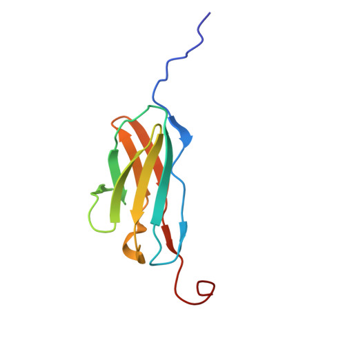
β-sandwich domains of the intermediate immunoglobulin fold (IgI) are key building blocks of many large, multidomain proteins - such as titin. The uniformity of the basic fold of these domains does not seem to be an impediment to their adaptation to a wide variety of functions. IgI domains in different regions of titin have to fulfil distinct functions which is seen in subtle differences of sequence, structure and stability. This is particularly true for the N2A region whose IgI domains are subtly distinct from other parts of I-band titin. We have already shown the unusual structure and properties of IgI domain I83 which is able to bind to calcium and is part of a binding site for F-actin and p94. To continue our exploration of the titin IgI domains of the N2A region we describe here the solution NMR structure of domain I82 of murine titin. The structure of the murine domain is virtually identical to the human homologue. However, the NMR investigation reveals the existence of distinct conformers around a highly conserved glycine, part of the tyrosine corner motif at the junction of the EF-loop with the F-strand. 15 N relaxation data show substantial line broadening for residues around this glycine, confirming conformational exchange on the fast-intermediate time scale. The unusual dynamics could for the first time explain the high level of conservation of the tyrosine corner glycine via a function in the folding of the domain.
- Chemistry Department, University of Massachusetts Lowell, Lowell, MA, 01854, USA.
Organizational Affiliation: