Structural Insights and Functional Dynamics of beta-Lactoglobulin Fibrils.
Sternke-Hoffmann, R., Rhyner, D., Terashi, G., Qureshi, B.M., Riek, R., Greenwald, J., Kihara, D., Lutz-Bueno, V., Luo, J.(2025) Nano Lett 25: 16146-16153
- PubMed: 41129746 Search on PubMed
- DOI: https://doi.org/10.1021/acs.nanolett.5c04125
- Primary Citation Related Structures:
9IAH - PubMed Abstract:
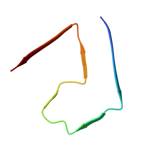
Amyloid fibrils from β-lactoglobulin (β-LG), a major whey protein, have attracted interest for nanotechnology due to their biocompatibility, tunable surface chemistry, and ability to bind functional molecules. They serve as scaffolds for metal nanoparticle synthesis, carriers for bioactive compounds, and building blocks for nanomaterials with tailored mechanical and optical properties. However, their dynamic architecture remains incompletely understood, limiting their rational design. Here, we combine cryo-electron microscopy (cryo-EM), small-angle X-ray scattering (SAXS), and molecular dynamics (MD) simulations to investigate β-LG fibrils formed under mildly denaturing conditions. Cryo-EM reveals a monomeric polymorph with a conserved core (Leu1-Ala34) and a disordered "fuzzy coat". Flexible domains were modeled and evaluated by MD, identifying one stable conformation (Asn90-Thr97). The ionic strength reduced the coat flexibility and promoted iron binding, suggesting environmental responsiveness. These findings link fibril flexibility to functional potential, offering mechanistic insight into engineering β-LG-based nanomaterials.
- PSI Center for Life Sciences, Paul Scherrer Institute, Forschungsstrasse 111, CH-5232 Villigen, PSI, Switzerland.
Organizational Affiliation: