Cryo-EM identifies F-ENA of Bacillus thuringiensis as a widespread family of endospore appendages across Firmicutes.
Sleutel, M., Sogues, A., Van Gerven, N., Jonsmoen, U.L., Van Molle, I., Fislage, M., Theunissen, L.D., Bellis, N.F., Baquero, D.P., Egelman, E.H., Krupovic, M., Wang, F., Aspholm, M., Remaut, H.(2025) Nat Commun 16: 7652-7652
- PubMed: 40818982 Search on PubMedSearch on PubMed Central
- DOI: https://doi.org/10.1038/s41467-025-62896-3
- Primary Citation Related Structures:
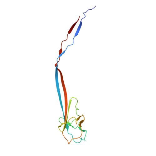
9HZE, 9I65, 9N0B - PubMed Abstract:
For over 100 years, Bacillus thuringiensis (Bt) has been used as an agricultural biopesticide to control pests caused by insect species in the orders of Lepidoptera, Diptera, and Coleoptera. Under nutrient starvation, Bt cells differentiate into spores and associated toxin crystals that can adopt biofilm-like aggregates. We reveal that such Bt spore/toxin biofilms are embedded in a fibrous extrasporal matrix, and using cryoID, we resolved the structure and molecular identity of an uncharacterized type of pili, referred to here as Fibrillar ENdospore Appendages or F-ENA. F-ENA are monomolecular protein filaments anchored to the exosporium and tipped with a flexible fibrillum. Phylogenetic and structural analyses reveal that F-ENA are conserved in Bacilli and Clostridia, featuring head-neck domains with β-barrel necks that interlock via N-terminal hook peptides. In Bacillus, two collagen-like proteins (F-Anchor and F-BclA), respectively, tether F-ENA and form the distal tip. Sedimentation assays suggest F-ENA promotes spore clustering via F-BclA contacts and/or filament bundling.
- Structural Biology Brussels, Vrije Universiteit Brussel, Brussels, Belgium. Mike.Sleutel@vub.be.
Organizational Affiliation: