9I4Z | pdb_00009i4z

Crystal structure of Thomasclavelia ramosa IgA peptidase (IgAse) active site mutant (E330-N876)


Experimental Data Snapshot

  • Method: X-RAY DIFFRACTION
  • Resolution: 1.75 Å
  • R-Value Free: 
    0.213 (Depositor), 0.199 (DCC) 
  • R-Value Work: 
    0.178 (Depositor), 0.171 (DCC) 
  • R-Value Observed: 
    0.178 (Depositor) 

Starting Model: in silico
View more details

wwPDB Validation   3D Report Full Report


This is version 2.0 of the entry. See complete history


Literature

Biochemical and structural characterization of the human gut microbiome metallopeptidase IgAse provides insight into its unique specificity for the Fab' region of IgA1 and IgA2.

Ramirez-Larrota, J.S.Juyoux, P.Guerra, P.Eckhard, U.Gomis-Ruth, F.X.

(2025) PLoS Pathog 21: e1013292-e1013292

  • DOI: https://doi.org/10.1371/journal.ppat.1013292
  • Primary Citation of Related Structures:  
    9I4Z, 9QA6

  • PubMed Abstract: 

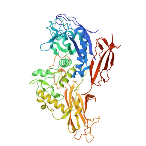
    Human immunoglobulin A (IgA), comprising the isotypes IgA1 and IgA2, protects ~400 m2 of mucosal surfaces against microbial infections but can also lead to aberrant IgA deposits that cause disease. Certain bacteria have evolved peptidases that cleave the hinge between the Fab and Fc fragments of IgA, undermining its immune function. These peptidases specifically target IgA1, but not IgA2, which predominates in the gut and possesses a structurally distinct hinge region. The only known IgA2-specific peptidase is IgAse from the gut microbiome member Thomasclavelia ramosa, which also targets IgA1 but no other proteins. IgAse is a ~ 140-kDa, seven-domain, membrane-bound metallopeptidase (MP). Differential scanning fluorimetry, small-angle X-ray scattering, AI-based structural predictions, mass spectrometry, and high-resolution crystallography and cryo-electron microscopy of multidomain fragments of IgAse revealed a novel 313-residue catalytic domain (CD) from the igalysin family within the metzincin MP clan. The CD is flanked by an N-terminal globular C-type lectin-like domain and a wrapping domain (WD), followed by four all-β domains. Functional studies involving a comprehensive set of constructs (wild-type and mutant), authentic and recombinant IgA fragments, and inhibitors demonstrated that the minimal functional assembly requires the CD and WD, along with the Fab and hinge region (Fab'). Modelling studies suggested that the Fab heavy-chain constant domain interacts with the N-terminal subdomain of the CD, positioning the hinge peptide for cleavage-a mechanism confirmed by mutational analysis. These findings open avenues for therapeutic strategies to inhibit the only known IgA1/IgA2 peptidase and to develop it for dissolving pathologic IgA deposits.


  • Organizational Affiliation
    • Proteolysis Laboratory, Department of Structural and Molecular Biology, Molecular Biology Institute of Barcelona (CSIC), Barcelona Science Park, Barcelona, Catalonia, Spain.

Macromolecules
Find similar proteins by:  (by identity cutoff)  |  3D Structure
Entity ID: 1
MoleculeChains Sequence LengthOrganismDetailsImage
IgA protease559Thomasclavelia ramosaMutation(s): 1 
Gene Names: iga
UniProt
Find proteins for Q9AES2 (Thomasclavelia ramosa)
Explore Q9AES2 
Go to UniProtKB:  Q9AES2
Entity Groups  
Sequence Clusters30% Identity50% Identity70% Identity90% Identity95% Identity100% Identity
UniProt GroupQ9AES2
Sequence Annotations
Expand
  • Reference Sequence
Small Molecules
Ligands 4 Unique
IDChains Name / Formula / InChI Key2D Diagram3D Interactions
PG4
Query on PG4

Download Ideal Coordinates CCD File 
D [auth A]TETRAETHYLENE GLYCOL
C8 H18 O5
UWHCKJMYHZGTIT-UHFFFAOYSA-N
ZN (Subject of Investigation/LOI)
Query on ZN

Download Ideal Coordinates CCD File 
B [auth A],
C [auth A]
ZINC ION
Zn
PTFCDOFLOPIGGS-UHFFFAOYSA-N
EDO
Query on EDO

Download Ideal Coordinates CCD File 
E [auth A]
F [auth A]
G [auth A]
H [auth A]
I [auth A]
E [auth A],
F [auth A],
G [auth A],
H [auth A],
I [auth A],
J [auth A],
K [auth A],
L [auth A]
1,2-ETHANEDIOL
C2 H6 O2
LYCAIKOWRPUZTN-UHFFFAOYSA-N
FMT
Query on FMT

Download Ideal Coordinates CCD File 
M [auth A]FORMIC ACID
C H2 O2
BDAGIHXWWSANSR-UHFFFAOYSA-N
Experimental Data & Validation

Experimental Data

  • Method: X-RAY DIFFRACTION
  • Resolution: 1.75 Å
  • R-Value Free:  0.213 (Depositor), 0.199 (DCC) 
  • R-Value Work:  0.178 (Depositor), 0.171 (DCC) 
  • R-Value Observed: 0.178 (Depositor) 
Space Group: P 1 21 1
Unit Cell:
Length ( Å )Angle ( ˚ )
a = 46.15α = 90
b = 87.83β = 96.11
c = 67.47γ = 90
Software Package:
Software NamePurpose
BUSTERrefinement
SHELXEmodel building
XDSdata reduction
XSCALEdata scaling
PHASERphasing

Structure Validation

View Full Validation Report


Entry History & Funding Information

Deposition Data


Funding OrganizationLocationGrant Number
Agencia Estatal de Investigacion (AEI)SpainPRE2020-096731
Agencia Estatal de Investigacion (AEI)SpainPID2019-107725RB-I00
Agencia Estatal de Investigacion (AEI)SpainRYC2020-029773-I

Revision History  (Full details and data files)

  • Version 1.0: 2025-06-25
    Type: Initial release
  • Version 1.1: 2025-07-02
    Changes: Structure summary
  • Version 2.0: 2025-07-23
    Changes: Atomic model, Data collection, Database references, Refinement description