9I1Q | pdb_00009i1q

HER3 receptor in complex with the Fab fragment of TK-hu A3 monoclonal antibody


Experimental Data Snapshot

  • Method: X-RAY DIFFRACTION
  • Resolution: 2.80 Å
  • R-Value Free: 
    0.263 (Depositor), 0.258 (DCC) 
  • R-Value Work: 
    0.209 (Depositor), 0.208 (DCC) 
  • R-Value Observed: 
    0.212 (Depositor) 

Starting Models: in silico, experimental
View more details

wwPDB Validation 3D Report Full Report

Validation slider image for 9I1Q

Ligand Structure Quality Assessment 


This is version 1.1 of the entry. See complete history

Literature

Novel Humanized Anti-HER3 Antibodies: Structural Characterization and Therapeutic Activity.

Muzi, A.Arriga, R.Bulfaro, G.Fata, F.Costanzo, A.Chiarini, V.Cappelletti, M.Ferrara, F.F.Bucci, F.Montemiglio, L.C.Savino, C.Marra, E.Ciliberto, G.Aurisicchio, L.Vallone, B.Roscilli, G.

(2025) Antibodies (Basel) 14

  • DOI: https://doi.org/10.3390/antib14040084
  • Primary Citation Related Structures: 
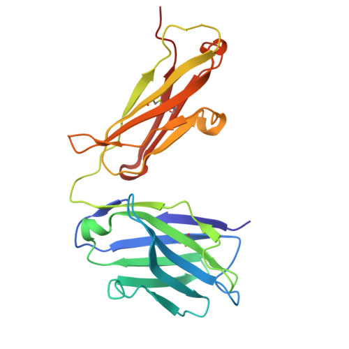
    9I1Q

  • PubMed Abstract: 

    The ErbB protein family plays a critical role in the progression of various solid tumors, and HER3 has been implicated in resistance mechanisms to multiple cancer therapies due to its ability to form heterodimers with other ErbB receptors, thereby activating pathways that promote tumor growth and survival. This study aimed to generate and characterize humanized monoclonal antibodies against HER3 to inhibit its function and evaluate their potential as therapeutic agents. Murine monoclonal antibodies TK-A3 and TK-A4 were humanized and tested for binding to ErbB3 and competition with neuregulin-1β (NRG). Specificity was assessed by ELISA, and epitope identified by X-ray crystallography. Downstream signaling was analyzed by western blot for phosphorylated ErbB3, Akt, and MAPK. Antitumor activity was evaluated in vitro and in a pancreatic cancer xenograft model. A toxicology study was also conducted. TK-hu A3 and TK-hu A4 bound specifically to ErbB3 without cross-reactivity to other ErbB receptors. The ErbB3-TK-hu A3 Fab structure revealed the binding epitope. Both antibodies competed with NRG, inhibiting ErbB3, Akt, and MAPK phosphorylation in a dose-dependent manner. They suppressed cancer cell survival in vitro, and TK-hu A3 significantly delayed tumor growth in vivo. The toxicology study indicated good tolerability. TK-hu A3 emerged as the lead candidate, showing specific HER3 targeting, strong pathway inhibition, and antitumor efficacy in vivo. Beyond standalone use, it could support novel strategies such as T-cell engagers, ADCs, CAR-T, and bispecific antibodies. These findings highlight TK-hu A3 as a promising therapy for HER3-positive, treatment-resistant cancers, meriting further development.


  • Organizational Affiliation
    • Takis s.r.l., 00128 Rome, Italy.

Macromolecule Content 

  • Total Structure Weight: 481 kDa 
  • Atom Count: 31,872 
  • Modeled Residue Count: 4,062 
  • Deposited Residue Count: 4,300 
  • Unique protein chains: 3

Macromolecules

Find similar proteins by:|  3D Structure
Entity ID: 1
MoleculeChains  Sequence LengthOrganismDetailsImage
Receptor tyrosine-protein kinase erbB-3
A, B, C, D
632Homo sapiensMutation(s): 0 
Gene Names: ERBB3HER3
EC: 2.7.10.1
UniProt & NIH Common Fund Data Resources
Find proteins for P21860 (Homo sapiens)
Explore P21860 
Go to UniProtKB:  P21860
PHAROS:  P21860
GTEx:  ENSG00000065361 
Entity Groups
Sequence Clusters30% Identity50% Identity70% Identity90% Identity95% Identity100% Identity
UniProt GroupP21860
Glycosylation
Glycosylation Sites: 8Go to GlyGen: P21860-1
Sequence Annotations
Expand
Reference Sequence
Find similar proteins by:|  3D Structure
Entity ID: 2
MoleculeChains  Sequence LengthOrganismDetailsImage
light chain hA3-Fab
E, F, G, H
220Homo sapiensMutation(s): 0 
Find similar proteins by:|  3D Structure
Entity ID: 3
MoleculeChains  Sequence LengthOrganismDetailsImage
heavy chain hA3-Fab
I, J, K, L
223Homo sapiensMutation(s): 0 

Oligosaccharides

Help  
Entity ID: 4
MoleculeChains Length2D Diagram GlycosylationD Interactions
beta-D-mannopyranose-(1-4)-2-acetamido-2-deoxy-beta-D-glucopyranose-(1-4)-2-acetamido-2-deoxy-beta-D-glucopyranose
M, O
3N-Glycosylation
Glycosylation Resources
GlyTouCan: G15407YE
GlyCosmos: G15407YE
GlyGen: G15407YE
Entity ID: 5
MoleculeChains Length2D Diagram GlycosylationD Interactions
alpha-D-mannopyranose-(1-6)-beta-D-mannopyranose-(1-4)-2-acetamido-2-deoxy-beta-D-glucopyranose-(1-4)-2-acetamido-2-deoxy-beta-D-glucopyranose
N
4N-Glycosylation
Glycosylation Resources
GlyTouCan: G22573RC
GlyCosmos: G22573RC
GlyGen: G22573RC
Entity ID: 6
MoleculeChains Length2D Diagram GlycosylationD Interactions
2-acetamido-2-deoxy-beta-D-glucopyranose-(1-4)-2-acetamido-2-deoxy-beta-D-glucopyranose
P, Q
2N-Glycosylation
Glycosylation Resources
GlyTouCan: G42666HT
GlyCosmos: G42666HT
GlyGen: G42666HT

Small Molecules

Ligands 2 Unique
IDChains Name / Formula / InChI Key2D Diagram3D Interactions
NAG

Query on NAG


Download:Ideal Coordinates CCD File
AA [auth B]
BA [auth B]
CA [auth B]
EA [auth C]
FA [auth C]
AA [auth B],
BA [auth B],
CA [auth B],
EA [auth C],
FA [auth C],
GA [auth C],
HA [auth C],
IA [auth C],
KA [auth D],
LA [auth D],
MA [auth D],
NA [auth D],
OA [auth D],
PA [auth D],
QA [auth D],
R [auth A],
S [auth A],
T [auth A],
U [auth A],
V [auth A],
W [auth A],
X [auth B],
Y [auth B],
Z [auth B]
2-acetamido-2-deoxy-beta-D-glucopyranose
C8 H15 N O6
OVRNDRQMDRJTHS-FMDGEEDCSA-N
SCN

Query on SCN


Download:Ideal Coordinates CCD File
DA [auth B]
JA [auth C]
RA [auth D]
SA [auth D]
TA [auth H]
DA [auth B],
JA [auth C],
RA [auth D],
SA [auth D],
TA [auth H],
UA [auth H],
VA [auth I],
WA [auth I],
XA [auth L]
THIOCYANATE ION
C N S
ZMZDMBWJUHKJPS-UHFFFAOYSA-M

Experimental Data & Validation

Experimental Data

  • Method: X-RAY DIFFRACTION
  • Resolution: 2.80 Å
  • R-Value Free:  0.263 (Depositor), 0.258 (DCC) 
  • R-Value Work:  0.209 (Depositor), 0.208 (DCC) 
  • R-Value Observed: 0.212 (Depositor) 
Space Group: P 1 21 1
Unit Cell:
Length ( Å )Angle ( ˚ )
a = 98.03α = 90
b = 98.81β = 99.21
c = 270.38γ = 90
Software Package:
Software NamePurpose
PHENIXrefinement
autoPROCdata reduction
autoPROCdata scaling
PHASERphasing

Structure Validation

View Full Validation Report


Ligand Structure Quality Assessment 


Entry History 

& Funding Information

Deposition Data


Funding OrganizationLocationGrant Number
Regione Lazio (Italy)Italy--

Revision History  (Full details and data files)

  • Version 1.0: 2025-10-15
    Type: Initial release
  • Version 1.1: 2025-11-05
    Changes: Database references