9I1J | pdb_00009i1j

Cryo-EM structure of mouse RNF213:UBE2L3 transthiolation intermediate, chemically stabilized, and ATPgS


Experimental Data Snapshot

  • Method: ELECTRON MICROSCOPY
  • Resolution: 3.80 Å
  • Aggregation State: PARTICLE 
  • Reconstruction Method: SINGLE PARTICLE 

wwPDB Validation   3D Report Full Report


This is version 1.0 of the entry. See complete history


Literature

ATP functions as a pathogen-associated molecular pattern to activate the E3 ubiquitin ligase RNF213.

Ahel, J.Balci, A.Faas, V.Grabarczyk, D.B.Harmo, R.Squair, D.R.Zhang, J.Roitinger, E.Lamoliatte, F.Mathur, S.Deszcz, L.Bell, L.E.Lehner, A.Williams, T.L.Sowar, H.Meinhart, A.Wood, N.T.Clausen, T.Virdee, S.Fletcher, A.J.

(2025) Nat Commun 16: 4414-4414

  • DOI: https://doi.org/10.1038/s41467-025-59444-4
  • Primary Citation of Related Structures:  
    9I1I, 9I1J

  • PubMed Abstract: 

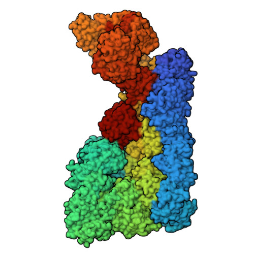
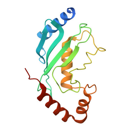
    The giant E3 ubiquitin ligase RNF213 is a conserved component of mammalian cell-autonomous immunity, limiting the replication of bacteria, viruses and parasites. To understand how RNF213 reacts to these unrelated pathogens, we employ chemical and structural biology to find that ATP binding to its ATPases Associated with diverse cellular Activities (AAA) core activates its E3 function. We develop methodology for proteome-wide E3 activity profiling inside living cells, revealing that RNF213 undergoes a reversible switch in E3 activity in response to cellular ATP abundance. Interferon stimulation of macrophages raises intracellular ATP levels and primes RNF213 E3 activity, while glycolysis inhibition depletes ATP and downregulates E3 activity. These data imply that ATP bears hallmarks of a danger/pathogen associated molecular pattern, coordinating cell-autonomous defence. Furthermore, quantitative labelling of RNF213 with E3-activity probes enabled us to identify the catalytic cysteine required for substrate ubiquitination and obtain a cryo-EM structure of the RNF213-E2-ubiquitin conjugation enzyme transfer intermediate, illuminating an unannotated E2 docking site. Together, our data demonstrate that RNF213 represents a new class of ATP-dependent E3 enzyme, employing distinct catalytic and regulatory mechanisms adapted to its specialised role in the broad defence against intracellular pathogens.


  • Organizational Affiliation
    • Research Institute of Molecular Pathology (IMP), Vienna BioCenter, Vienna, Austria.

Macromolecules
Find similar proteins by:  (by identity cutoff)  |  3D Structure
Entity ID: 1
MoleculeChains Sequence LengthOrganismDetailsImage
E3 ubiquitin-protein ligase RNF2134,836Mus musculusMutation(s): 0 
Gene Names: Rnf213Mystr
EC: 2.3.2.27 (PDB Primary Data), 3.6.4 (PDB Primary Data), 2.3.2 (UniProt)
UniProt
Find proteins for E9Q555 (Mus musculus)
Explore E9Q555 
Go to UniProtKB:  E9Q555
Entity Groups  
Sequence Clusters30% Identity50% Identity70% Identity90% Identity95% Identity100% Identity
UniProt GroupE9Q555
Sequence Annotations
Expand
  • Reference Sequence
Find similar proteins by:  (by identity cutoff)  |  3D Structure
Entity ID: 2
MoleculeChains Sequence LengthOrganismDetailsImage
Ubiquitin-conjugating enzyme E2 L3166Homo sapiensMutation(s): 0 
Gene Names: UBE2L3UBCE7UBCH7
EC: 2.3.2.23
UniProt & NIH Common Fund Data Resources
Find proteins for P68036 (Homo sapiens)
Explore P68036 
Go to UniProtKB:  P68036
PHAROS:  P68036
GTEx:  ENSG00000185651 
Entity Groups  
Sequence Clusters30% Identity50% Identity70% Identity90% Identity95% Identity100% Identity
UniProt GroupP68036
Sequence Annotations
Expand
  • Reference Sequence
Small Molecules
Ligands 4 Unique
IDChains Name / Formula / InChI Key2D Diagram3D Interactions
ATP (Subject of Investigation/LOI)
Query on ATP

Download Ideal Coordinates CCD File 
C [auth A]ADENOSINE-5'-TRIPHOSPHATE
C10 H16 N5 O13 P3
ZKHQWZAMYRWXGA-KQYNXXCUSA-N
ADP (Subject of Investigation/LOI)
Query on ADP

Download Ideal Coordinates CCD File 
G [auth A],
H [auth A]
ADENOSINE-5'-DIPHOSPHATE
C10 H15 N5 O10 P2
XTWYTFMLZFPYCI-KQYNXXCUSA-N
ZN
Query on ZN

Download Ideal Coordinates CCD File 
E [auth A],
F [auth A]
ZINC ION
Zn
PTFCDOFLOPIGGS-UHFFFAOYSA-N
MG (Subject of Investigation/LOI)
Query on MG

Download Ideal Coordinates CCD File 
D [auth A]MAGNESIUM ION
Mg
JLVVSXFLKOJNIY-UHFFFAOYSA-N
Experimental Data & Validation

Experimental Data

  • Method: ELECTRON MICROSCOPY
  • Resolution: 3.80 Å
  • Aggregation State: PARTICLE 
  • Reconstruction Method: SINGLE PARTICLE 
EM Software:
TaskSoftware PackageVersion
MODEL REFINEMENTPHENIX1.21.2_5419

Structure Validation

View Full Validation Report


Entry History & Funding Information

Deposition Data


Funding OrganizationLocationGrant Number
Austrian Research Promotion AgencyAustria852936

Revision History  (Full details and data files)

  • Version 1.0: 2025-12-10
    Type: Initial release