9I0O | pdb_00009i0o

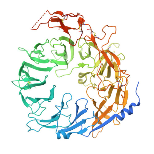
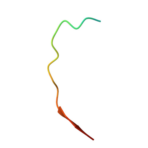
Cryo-EM structure of human sortilin ectodomain in complex with a thyroglobulin C-terminal peptide


Experimental Data Snapshot

  • Method: ELECTRON MICROSCOPY
  • Resolution: 3.36 Å
  • Aggregation State: PARTICLE 
  • Reconstruction Method: SINGLE PARTICLE 

Starting Model: experimental
View more details

wwPDB Validation 3D Report Full Report

Validation slider image for 9I0O

This is version 1.1 of the entry. See complete history

Literature

Molecular recognition of thyroglobulin by sortilin.

Boniardi, I.Tanzi, G.Di Ianni, A.Graziadei, A.Nedeljkovic, M.Stejskalova, C.Coscia, F.

(2026) Nat Commun 17

  • DOI: https://doi.org/10.1038/s41467-026-68658-z
  • Primary Citation Related Structures: 
    9I0N, 9I0O

  • PubMed Abstract: 

    Sortilin is a ubiquitous membrane receptor mediating trafficking of protein cargoes. In the thyroid, sortilin binds thyroglobulin (TG) during its endocytosis, a key process in thyroid homeostasis. Although sortilin has been proposed to recognise highly iodinated TG, the molecular details of this interaction remain unknown. In this work, using an integrative structural biology approach, we reveal that sortilin binds an unstructured TG C-terminal peptide and exhibits a strong preference for the monomeric TG over the commonly known dimeric form. We find that sortilin-TG interaction is independent of the iodination state of TG and instead relies on the conversion to its monomeric state, presumably promoted by extracellular degradation. Furthermore, using AlphaPulldown and sequence analysis, we show that recognition of other reported ligands by sortilin likely relies on similar unstructured peptide motifs, which are not constrained to a single binding orientation within the receptor cavity. Overall, this study reveals the TG-sortilin binding interface and provides insights into the recognition mechanism of other cargoes by sortilin.


  • Organizational Affiliation
    • Human Technopole, Milan, Italy.

Macromolecule Content 

  • Total Structure Weight: 86.22 kDa 
  • Atom Count: 5,054 
  • Modeled Residue Count: 636 
  • Deposited Residue Count: 762 
  • Unique protein chains: 2

Macromolecules

Find similar proteins by:|  3D Structure
Entity ID: 1
MoleculeChains  Sequence LengthOrganismDetailsImage
Sortilin742Homo sapiensMutation(s): 0 
Gene Names: SORT1
UniProt & NIH Common Fund Data Resources
Find proteins for Q99523 (Homo sapiens)
Explore Q99523 
Go to UniProtKB:  Q99523
PHAROS:  Q99523
GTEx:  ENSG00000134243 
Entity Groups
Sequence Clusters30% Identity50% Identity70% Identity90% Identity95% Identity100% Identity
UniProt GroupQ99523
Glycosylation
Glycosylation Sites: 3Go to GlyGen: Q99523-1
Sequence Annotations
Expand
Reference Sequence
Find similar proteins by:  Sequence   |   3D Structure  
Entity ID: 2
MoleculeChains  Sequence LengthOrganismDetailsImage
Thyroglobulin20Homo sapiensMutation(s): 0 
UniProt & NIH Common Fund Data Resources
Find proteins for P01266 (Homo sapiens)
Explore P01266 
Go to UniProtKB:  P01266
PHAROS:  P01266
GTEx:  ENSG00000042832 
Entity Groups
Sequence Clusters30% Identity50% Identity70% Identity90% Identity95% Identity100% Identity
UniProt GroupP01266
Sequence Annotations
Expand
Reference Sequence

Oligosaccharides

Help  
Entity ID: 3
MoleculeChains Length2D Diagram GlycosylationD Interactions
2-acetamido-2-deoxy-beta-D-glucopyranose-(1-4)-2-acetamido-2-deoxy-beta-D-glucopyranose
C, D
2N-Glycosylation
Glycosylation Resources
GlyTouCan: G42666HT
GlyCosmos: G42666HT
GlyGen: G42666HT

Small Molecules

Ligands 1 Unique
IDChains Name / Formula / InChI Key2D Diagram3D Interactions
NAG

Query on NAG


Download:Ideal Coordinates CCD File
E [auth A]2-acetamido-2-deoxy-beta-D-glucopyranose
C8 H15 N O6
OVRNDRQMDRJTHS-FMDGEEDCSA-N

Experimental Data & Validation

Experimental Data

  • Method: ELECTRON MICROSCOPY
  • Resolution: 3.36 Å
  • Aggregation State: PARTICLE 
  • Reconstruction Method: SINGLE PARTICLE 
EM Software:
TaskSoftware PackageVersion
MODEL REFINEMENTPHENIX1.21.1_5286

Structure Validation

View Full Validation Report


Entry History 

& Funding Information

Deposition Data


Funding OrganizationLocationGrant Number
European Research Council (ERC)European Union101041298

Revision History  (Full details and data files)

  • Version 1.0: 2025-12-17
    Type: Initial release
  • Version 1.1: 2026-07-01
    Changes: Data collection, Database references