9I0M | pdb_00009i0m

Mycobacterium smegmatis inosine monophosphate dehydrogenase (IMPDH) saturating ATP+IMP-bound form, extended


Experimental Data Snapshot

  • Method: ELECTRON MICROSCOPY
  • Resolution: 3.01 Å
  • Aggregation State: PARTICLE 
  • Reconstruction Method: SINGLE PARTICLE 

Starting Model: experimental
View more details

wwPDB Validation   3D Report Full Report


This is version 1.1 of the entry. See complete history


Literature

Conformational landscape of the mycobacterial inosine 5'-monophosphate dehydrogenase octamerization interface.

Bulvas, O.Knejzlik, Z.Filimonenko, A.Kouba, T.Pichova, I.

(2025) J Struct Biol 217: 108198-108198

  • DOI: https://doi.org/10.1016/j.jsb.2025.108198
  • Primary Citation of Related Structures:  
    9I0K, 9I0L, 9I0M

  • PubMed Abstract: 

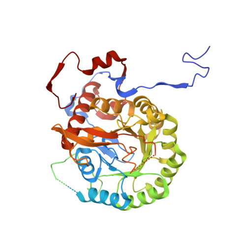
    Inosine 5'-monophosphate dehydrogenase (IMPDH), a key enzyme in bacterial purine metabolism, plays an essential role in the biosynthesis of guanine nucleotides and shows promise as a target for antimicrobial drug development. Despite its significance, the conformational dynamics and substrate-induced structural changes in bacterial IMPDH remain poorly understood, particularly with respect to its octameric assembly. Using cryo-EM, we present full-length structures of IMPDH from Mycobacterium smegmatis (MsmIMPDH) captured in a reaction intermediate state, revealing conformational changes upon substrate binding. The structures feature resolved flexible loops that coordinate the binding of the substrate, the cofactor, and the K + ion. Our structural analysis identifies a novel octamerization interface unique to MsmIMPDH. Additionally, a previously unobserved barrel-like density suggests potential self-interactions within the C-terminal regions, hinting at a regulatory mechanism tied to assembly and function of the enzyme. These data provide insights into substrate-induced conformational dynamics and novel interaction interfaces in MsmIMPDH, potentially informing the development of IMPDH-targeted drugs.


  • Organizational Affiliation
    • Institute of Organic Chemistry and Biochemistry of the Czech Academy of Sciences, Prague, Czech Republic.

Macromolecules
Find similar proteins by:  (by identity cutoff)  |  3D Structure
Entity ID: 1
MoleculeChains Sequence LengthOrganismDetailsImage
Inosine-5'-monophosphate dehydrogenase
A, B, C, D, E
A, B, C, D, E, F, G, H
513Mycolicibacterium smegmatis MC2 155Mutation(s): 0 
Gene Names: guaBMSMEG_1602
EC: 1.1.1.205
UniProt
Find proteins for P9WKI7 (Mycobacterium tuberculosis (strain ATCC 25618 / H37Rv))
Explore P9WKI7 
Go to UniProtKB:  P9WKI7
Entity Groups  
Sequence Clusters30% Identity50% Identity70% Identity90% Identity95% Identity100% Identity
UniProt GroupP9WKI7
Sequence Annotations
Expand
  • Reference Sequence
Small Molecules
Ligands 2 Unique
IDChains Name / Formula / InChI Key2D Diagram3D Interactions
IMP
Query on IMP

Download Ideal Coordinates CCD File 
I [auth A]
K [auth B]
M [auth C]
O [auth D]
Q [auth E]
I [auth A],
K [auth B],
M [auth C],
O [auth D],
Q [auth E],
S [auth F],
U [auth G],
X [auth H]
INOSINIC ACID
C10 H13 N4 O8 P
GRSZFWQUAKGDAV-KQYNXXCUSA-N
K
Query on K

Download Ideal Coordinates CCD File 
J [auth B]
L [auth B]
N [auth C]
P [auth D]
R [auth E]
J [auth B],
L [auth B],
N [auth C],
P [auth D],
R [auth E],
T [auth F],
V [auth G],
W [auth G]
POTASSIUM ION
K
NPYPAHLBTDXSSS-UHFFFAOYSA-N
Experimental Data & Validation

Experimental Data

  • Method: ELECTRON MICROSCOPY
  • Resolution: 3.01 Å
  • Aggregation State: PARTICLE 
  • Reconstruction Method: SINGLE PARTICLE 
EM Software:
TaskSoftware PackageVersion
MODEL REFINEMENTPHENIX1.21rc1_5084

Structure Validation

View Full Validation Report


Entry History & Funding Information

Deposition Data


Funding OrganizationLocationGrant Number
European Union (EU)European UnionLX22NPO5103

Revision History  (Full details and data files)

  • Version 1.0: 2025-03-26
    Type: Initial release
  • Version 1.1: 2025-04-02
    Changes: Data collection, Database references