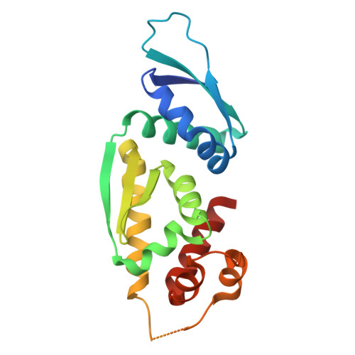
Structural and functional characterization of the extended-diKH domain from the antiviral endoribonuclease KHNYN.
Youle, R.L., Lista, M.J., Bouton, C., Kunzelmann, S., Wilson, H., Cottee, M.A., Purkiss, A.G., Morris, E.R., Neil, S.J.D., Taylor, I.A., Swanson, C.M.(2025) J Biological Chem 301: 108336-108336
- PubMed: 39984050 Search on PubMedSearch on PubMed Central
- DOI: https://doi.org/10.1016/j.jbc.2025.108336
- Primary Citation Related Structures:
9HTS - PubMed Abstract:
Zinc finger antiviral protein (ZAP) binds CpG dinucleotides in viral RNA and targets them for decay. ZAP interacts with several cofactors to form the ZAP antiviral system, including KHNYN, a multidomain endoribonuclease required for ZAP-mediated RNA decay. However, it is unclear how the individual domains in KHNYN contribute to its activity. Here, we demonstrate that the KHNYN amino-terminal extended-diKH (ex-diKH) domain is required for antiviral activity and present its crystal structure. The structure belongs to a rare group of KH-containing domains, characterized by a noncanonical arrangement between two type 1 KH modules, with an additional helical bundle. N4BP1 is a KHNYN paralog with an ex-diKH domain that functionally complements the KHNYN ex-diKH domain. Interestingly, the ex-diKH domain structure is present in N4BP1-like proteins in lancelets, which are basal chordates, indicating that it is evolutionarily ancient. While many KH domains demonstrate RNA binding activity, biolayer interferometry and electrophoretic mobility shift assays indicate that the KHNYN ex-diKH domain does not bind RNA. Furthermore, residues required for canonical KH domains to bind RNA are not required for KHNYN antiviral activity. By contrast, an inter-KH domain cleft in KHNYN is a potential protein-protein interaction site, and mutations that eliminate arginine salt bridges at the edge of this cleft decrease KHNYN antiviral activity. This suggests that this domain could be a binding site for an unknown KHNYN cofactor.
- Department of Infectious Diseases, King's College London, London, United Kingdom; Macromolecular Structure Laboratory, The Francis Crick Institute, London, United Kingdom.
Organizational Affiliation: