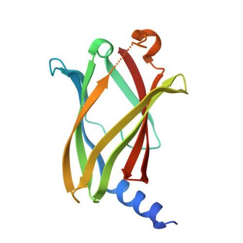
9HMC | pdb_00009hmc

Crystal structure of PDE6D in complex with compound 5e


Experimental Data Snapshot

  • Method: X-RAY DIFFRACTION
  • Resolution: 1.65 Å
  • R-Value Free: 
    0.223 (Depositor), 0.226 (DCC) 
  • R-Value Work: 
    0.188 (Depositor), 0.190 (DCC) 
  • R-Value Observed: 
    0.190 (Depositor) 

Starting Model: experimental
View more details

wwPDB Validation 3D Report Full Report

Validation slider image for 9HMC

Ligand Structure Quality Assessment 


This is version 1.1 of the entry. See complete history

Literature

Covalent modification of a glutamic acid inspired by HaloTag technology.

Zhang, R.Liu, J.Gasper, R.Unger, A.Kaschani, F.Kaiser, M.Janning, P.Waldmann, H.

(2026) Nat Commun 17: 1257-1257

  • DOI: https://doi.org/10.1038/s41467-026-68999-9
  • Primary Citation Related Structures: 
    9HMC, 9HMD

  • PubMed Abstract: 

    For targeted covalent protein modification at low-reactivity aspartates and glutamates, new methods are in high demand. We report a technique inspired by the HaloTag technology, which employs nucleophilic substitution at chloroalkane-functionalised ligands by a specific aspartate residue. Embedding of alkyl bromide warheads into non-covalent inhibitors enables covalent modification of a glutamate in the lipoprotein binding chaperone - phosphodiesterase of retinal rod subunit delta (PDEδ), which shuttles prenylated lipoproteins between cellular membranes and thereby mediates their activity. Its hydrophobic ligand-binding pocket contains p.E88 as the only accessible nucleophile for covalent targeting. We show that a covalent inhibitor, termed DeltaTag, overcomes limitations of non-covalent inhibitors. DeltaTag labels PDEδ at its p.E88 under biologically relevant conditions, modulates mammalian target of rapamycin (mTOR) signalling by disrupting the PDEδ-Rheb (Ras homologue enriched in brain)-mTORC1 (mTOR complex 1) axis and inhibits cancer cell proliferation. This proof-of-concept study demonstrates that the design strategy holds promise for the covalent modification of proteins with lipophilic binding sites that lack accessible reactive amino acids but contain specific carboxylates.


  • Organizational Affiliation
    • Department of Chemical Biology, Max Planck Institute of Molecular Physiology, Dortmund, Germany.

Macromolecule Content 

  • Total Structure Weight: 38 kDa 
  • Atom Count: 2,704 
  • Modeled Residue Count: 297 
  • Deposited Residue Count: 312 
  • Unique protein chains: 1

Macromolecules

Find similar proteins by:|  3D Structure
Entity ID: 1
MoleculeChains  Sequence LengthOrganismDetailsImage
Retinal rod rhodopsin-sensitive cGMP 3',5'-cyclic phosphodiesterase subunit delta
A, B
156Homo sapiensMutation(s): 0 
Gene Names: PDE6DPDED
UniProt & NIH Common Fund Data Resources
Find proteins for O43924 (Homo sapiens)
Explore O43924 
Go to UniProtKB:  O43924
PHAROS:  O43924
GTEx:  ENSG00000156973 
Entity Groups
Sequence Clusters30% Identity50% Identity70% Identity90% Identity95% Identity100% Identity
UniProt GroupO43924
Sequence Annotations
Expand
Reference Sequence

Experimental Data & Validation

Experimental Data

  • Method: X-RAY DIFFRACTION
  • Resolution: 1.65 Å
  • R-Value Free:  0.223 (Depositor), 0.226 (DCC) 
  • R-Value Work:  0.188 (Depositor), 0.190 (DCC) 
  • R-Value Observed: 0.190 (Depositor) 
Space Group: P 21 21 21
Unit Cell:
Length ( Å )Angle ( ˚ )
a = 56.74α = 90
b = 75.65β = 90
c = 82.31γ = 90
Software Package:
Software NamePurpose
PHENIXrefinement
XSCALEdata scaling
XDSdata reduction
PHASERphasing

Structure Validation

View Full Validation Report


Ligand Structure Quality Assessment 


Entry History 

& Funding Information

Deposition Data


Funding OrganizationLocationGrant Number
Not funded--

Revision History  (Full details and data files)

  • Version 1.0: 2025-12-24
    Type: Initial release
  • Version 1.1: 2026-07-08
    Changes: Database references