9HLB | pdb_00009hlb

TRPML1 in complex with compound 10


Experimental Data Snapshot

  • Method: ELECTRON MICROSCOPY
  • Resolution: 2.10 Å
  • Aggregation State: PARTICLE 
  • Reconstruction Method: SINGLE PARTICLE 

Starting Model: experimental
View more details

wwPDB Validation   3D Report Full Report


This is version 1.1 of the entry. See complete history


Literature

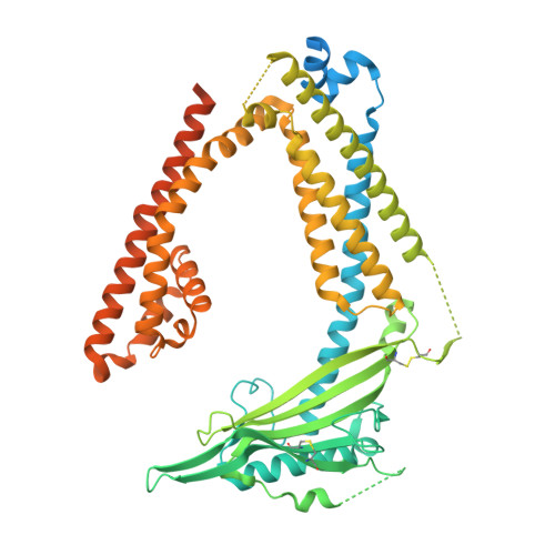
High throughput cryo-EM provides structural understanding for modulators of the lysosomal ion channel TRPML1.

Reeks, J.Mahajan, P.Clark, M.Cowan, S.R.Di Daniel, E.Earl, C.P.Fisher, S.Holvey, R.S.Jackson, S.M.Lloyd-Evans, E.Morgillo, C.M.Mortenson, P.N.O'Reilly, M.Richardson, C.J.Schopf, P.Tams, D.M.Waller-Evans, H.Ward, S.E.Whibley, S.Williams, P.A.Johnson, C.N.

(2025) Structure 33: 1374-1385.e7

  • DOI: https://doi.org/10.1016/j.str.2025.05.014
  • Primary Citation of Related Structures:  
    9HJ6, 9HJ8, 9HL3, 9HL4, 9HL6, 9HL8, 9HLA, 9HLB, 9HLC, 9HLD

  • PubMed Abstract: 

    Access to high-resolution structural data for protein-ligand complexes is a prerequisite for structure-based medicinal chemistry, where the ability to iterate cycles of design-structure-redesign is highly desirable. For proteins refractory to X-ray crystallography, such as integral membrane proteins, enablement of high throughput structure determination by cryoelectron microscopy (cryo-EM) has the potential to be transformational for structure-based design. We have applied such an approach to the lysosomal ion channel transient receptor potential mucolipin 1 (TRPML1) in complex with ten chemically diverse modulators, both agonists and antagonists. The resulting depth of high-resolution structural data generated provides important insights into protein-ligand structure-function relationships, including mechanistic understanding of ligand-induced channel pore opening and closing. Moreover, the knowledge gained has the potential to support iterative design cycles toward improved modulators of this important biological target.


  • Organizational Affiliation
    • Astex Pharmaceuticals, 436 Cambridge Science Park, Milton Road, Cambridge CB4 0QA, UK.

Macromolecules
Find similar proteins by:  (by identity cutoff)  |  3D Structure
Entity ID: 1
MoleculeChains Sequence LengthOrganismDetailsImage
Mucolipin-1A,
B [auth C],
C [auth B],
D
628Homo sapiensMutation(s): 0 
Gene Names: MCOLN1ML4TRPML1MSTP080
UniProt & NIH Common Fund Data Resources
Find proteins for Q9GZU1 (Homo sapiens)
Explore Q9GZU1 
Go to UniProtKB:  Q9GZU1
PHAROS:  Q9GZU1
GTEx:  ENSG00000090674 
Entity Groups  
Sequence Clusters30% Identity50% Identity70% Identity90% Identity95% Identity100% Identity
UniProt GroupQ9GZU1
Glycosylation
Glycosylation Sites: 1Go to GlyGen: Q9GZU1-1
Sequence Annotations
Expand
  • Reference Sequence
Oligosaccharides

Help

Entity ID: 2
MoleculeChains Length2D Diagram Glycosylation3D Interactions
2-acetamido-2-deoxy-beta-D-glucopyranose-(1-4)-2-acetamido-2-deoxy-beta-D-glucopyranose
E, F, G, H
2N-Glycosylation
Glycosylation Resources
GlyTouCan:  G42666HT
GlyCosmos:  G42666HT
GlyGen:  G42666HT
Small Molecules
Ligands 7 Unique
IDChains Name / Formula / InChI Key2D Diagram3D Interactions
EUJ
Query on EUJ

Download Ideal Coordinates CCD File 
LA [auth B],
N [auth A],
VA [auth D],
Y [auth C]
(2R)-3-{[(S)-hydroxy{[(1S,2R,3R,4S,5S,6R)-2,4,6-trihydroxy-3,5-bis(phosphonooxy)cyclohexyl]oxy}phosphoryl]oxy}propane-1,2-diyl dioctanoate
C25 H49 O19 P3
QXHVLVSULWMTCV-DICZBTHZSA-N
A1IV4 (Subject of Investigation/LOI)
Query on A1IV4

Download Ideal Coordinates CCD File 
EA [auth C],
GA [auth B],
QA [auth D],
T [auth A]
N-[2-[4-(2-methoxyphenyl)piperazin-1-yl]phenyl]benzenesulfonamide
C23 H25 N3 O3 S
DJQHXEVUPWVZGD-UHFFFAOYSA-N
R16
Query on R16

Download Ideal Coordinates CCD File 
MA [auth B],
O [auth A],
WA [auth D],
Z [auth C]
HEXADECANE
C16 H34
DCAYPVUWAIABOU-UHFFFAOYSA-N
D12
Query on D12

Download Ideal Coordinates CCD File 
AA [auth C]
DA [auth C]
FA [auth B]
IA [auth B]
J [auth A]
AA [auth C],
DA [auth C],
FA [auth B],
IA [auth B],
J [auth A],
K [auth A],
NA [auth B],
P [auth A],
S [auth A],
SA [auth D],
V [auth C],
XA [auth D]
DODECANE
C12 H26
SNRUBQQJIBEYMU-UHFFFAOYSA-N
D10
Query on D10

Download Ideal Coordinates CCD File 
BA [auth C],
OA [auth B],
Q [auth A],
YA [auth D]
DECANE
C10 H22
DIOQZVSQGTUSAI-UHFFFAOYSA-N
OCT
Query on OCT

Download Ideal Coordinates CCD File 
CA [auth C]
JA [auth B]
L [auth A]
PA [auth B]
R [auth A]
CA [auth C],
JA [auth B],
L [auth A],
PA [auth B],
R [auth A],
TA [auth D],
W [auth C],
ZA [auth D]
N-OCTANE
C8 H18
TVMXDCGIABBOFY-UHFFFAOYSA-N
HEX
Query on HEX

Download Ideal Coordinates CCD File 
HA [auth B]
I [auth A]
KA [auth B]
M [auth A]
RA [auth D]
HA [auth B],
I [auth A],
KA [auth B],
M [auth A],
RA [auth D],
U [auth C],
UA [auth D],
X [auth C]
HEXANE
C6 H14
VLKZOEOYAKHREP-UHFFFAOYSA-N
Experimental Data & Validation

Experimental Data

  • Method: ELECTRON MICROSCOPY
  • Resolution: 2.10 Å
  • Aggregation State: PARTICLE 
  • Reconstruction Method: SINGLE PARTICLE 
EM Software:
TaskSoftware PackageVersion
RECONSTRUCTIONcryoSPARC4
MODEL REFINEMENTPHENIX1.21rc1-5101

Structure Validation

View Full Validation Report


Entry History & Funding Information

Revision History  (Full details and data files)

  • Version 1.0: 2025-06-04
    Type: Initial release
  • Version 1.1: 2025-12-17
    Changes: Data collection, Database references