Unravelling the amyloid aggregation mechanism of the sweet protein Monellin: Insights from circular permutated mutants.
Lucignano, R., Bologna, A., Gramazio, S., Wang, P.H., Taxis, C., Essen, L.O., Picone, D., Spadaccini, R.(2025) Int J Biol Macromol 308: 142239-142239
- PubMed: 40118405 Search on PubMed
- DOI: https://doi.org/10.1016/j.ijbiomac.2025.142239
- Primary Citation Related Structures:
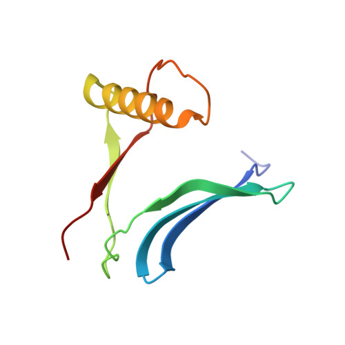
9HIJ, 9HIK, 9HKG - PubMed Abstract:
Protein amyloid aggregates, once regarded solely as pathological hallmarks of human neurodegenerative diseases, have recently gained attention for their potential in biotechnological applications. Among others, MNEI and its variants, initially developed as single-chain derivatives of the sweet protein monellin, also serve as valuable models for studying protein fibrillary aggregation. In this work, we have characterized three circular permutated mutants of MNEI obtained joining the N- and C-termini of MNEI with linkers of different length and restoring the splitting of the polypeptide chain of native monellin. All proteins are well folded but have a different propensity to form oligomeric structures in solution and aggregation rates comparable to or faster than MNEI, as indicated by Thioflavin-T binding assays. Transmission Electron Microscopy (TEM) studies indicate that only Perm1, the mutant with the longest linker, forms fibrillar aggregates. X-ray structures of the mutants show that they crystallize as domain-swapped dimers. Molecular dynamics study highlights potential hot spots controlling the ordered aggregation process of Perm1. Our data support the idea that the formation of a domain-swapped dimer does not favour the formation of fibrillar aggregates and highlight circular permutation as a valuable tool to build nanostructured biomaterials.
- Department of Chemical Sciences, University of Naples Federico II, Naples, Campania, Italy.
Organizational Affiliation: