Download Mendeley
Receptor-substrate competition for the TonB homolog FusB suggests a model for ferredoxin import.
Wojnowska, M., Flores, V., Yelland, T., Fisher, S.R., Bogucka, A., Stott, K., Walker, D.(2026) J Biol Chem 302: 113121-113121
- PubMed: 42103222 Search on PubMedSearch on PubMed Central
- DOI: https://doi.org/10.1016/j.jbc.2026.113121
- Primary Citation Related Structures:
7ZC8, 9HI3 - PubMed Abstract:
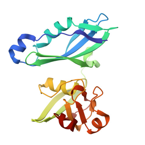
TonB-dependent uptake systems of gram-negative bacterial pathogens constitute prominent virulence factors, allowing nutrients-primarily siderophore-bound iron-to cross the highly impermeable outer membrane. Remarkably, the ferredoxin uptake system (Fus) of certain soft rot plant pathogens imports an entire host protein into the periplasm and extracts its bound iron for growth. The inner membrane protein FusB, a TonB homolog, plays two roles in facilitating import. First, like other TonBs, it remodels the globular plug domain obstructing the lumen of the outer membrane receptor FusA to allow ferredoxin passage. Unusually for a TonB protein, FusB then interacts directly with the FusA-bound ferredoxin substrate to facilitate its transport into the periplasm. Here, we describe structures of the FusB homodimer as well as the FusB-ferredoxin complex and, using biophysical, biochemical, and mutagenesis approaches, we determine the key features of the binding interfaces formed by FusB with FusA and ferredoxin. The C-terminal domain of FusB (FusB-CTD) exists in a monomer-dimer equilibrium in vitro, with the homodimer stabilized by an intermolecular R241-D322 salt bridge. The "FusB-box" of FusA interacts with monomeric FusB-CTD, and FusA D53 outcompetes FusB D322 to bind R241. Upon ferredoxin binding, FusB-CTD undergoes a structural rearrangement, expanding its β-sheet from three to four strands. In agreement with the proposed sequence of events, ferredoxin binding displaces FusA from FusB with R241 forming an intramolecular salt bridge with D322 to stabilize the newly formed β-hairpin of FusB. We propose a mechanistic model for ferredoxin import where FusB R241 acts as a molecular switch.
- Department of Biochemistry, University of Cambridge, Cambridge, United Kingdom. Electronic address: mw915@cam.ac.uk.
Organizational Affiliation: