A single residue affects the dynamics and shape of a tetrameric GH43 beta-1,4-d-xylosidase from Levilactobacillus brevis DSM1269.
Linares-Pasten, J.A., Faryar, R., Torrez Alvarez, S., Albasri, K., Shuoker, B., Abou Hachem, M., Logan, D.T., Karlsson, E.N.(2025) Protein Sci 34: e70299-e70299
- PubMed: 40944464 Search on PubMedSearch on PubMed Central
- DOI: https://doi.org/10.1002/pro.70299
- Primary Citation Related Structures:
9HE8 - PubMed Abstract:
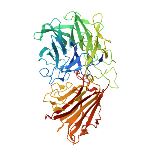
Lignocellulosic materials (e.g., straw and bran) are gaining interest as feedstocks for the manufacture of higher value products, recognizing xylooligosaccharides (XOS) as interesting prebiotic compounds, and putting enzymes converting xylooligosaccharides into focus. In this work, we are investigating a XOS converting enzyme from the probiotic bacterium Levilactobacillus brevis (formerly Lactobacillus brevis). Growth of multiple L. brevis strains, recognized as probiotics, is promoted by the presence of XOS. This study elucidates the 3D structure of an intracellular L. brevis β-xylosidase, LbXyn43B from glycoside hydrolase family 43 (GH43), resolving it to 1.9 Å resolution. Functional analysis identified LbXyn43B as a mediator for XOS utilization in L. brevis DSM1269. The enzyme, featuring a 5-fold β-propeller fold typical of GH43, prefers short XOS substrates, notably β-1,4-xylobiose and β-1,4-xylotriose, aligning with its role in hydrolyzing internalized XOS. Intriguingly, crystallographic and size exclusion chromatographic evidence reveals LbXyn43B to be a tetramer, contrary to the dimeric structure previously reported in a closely related homolog from strain DSM20054. Despite sharing a high sequence identity (differing in only five residues), Thr274 in LbXyn43B within a subunit interface loop was found to influence the tetramer's shape. When mutated to Ala (the residue at the corresponding position in the homolog), the enzyme's apparent native molecular mass was impacted, resulting in a more compact oligomeric structure of slightly higher thermostability. Kinetic analysis and molecular dynamics simulations further suggest an effect of Thr274Ala substitution in modulating the accessibility of longer oligosaccharides, such as β-1,4-d-xylotetraose, to the active site.
- Biotechnology and Applied Microbiology, Department of Process and Life Sciences Engineering, LTH, Lund University, Lund, Sweden.
Organizational Affiliation: