9HAP | pdb_00009hap

Cryo-EM structure of inactive human arginine-vasopressin (AVP) V2 receptor (V2R) with tolvaptan


Experimental Data Snapshot

  • Method: ELECTRON MICROSCOPY
  • Resolution: 2.50 Å
  • Aggregation State: PARTICLE 
  • Reconstruction Method: SINGLE PARTICLE 

Starting Model: in silico
View more details

wwPDB Validation   3D Report Full Report


This is version 1.0 of the entry. See complete history


Literature

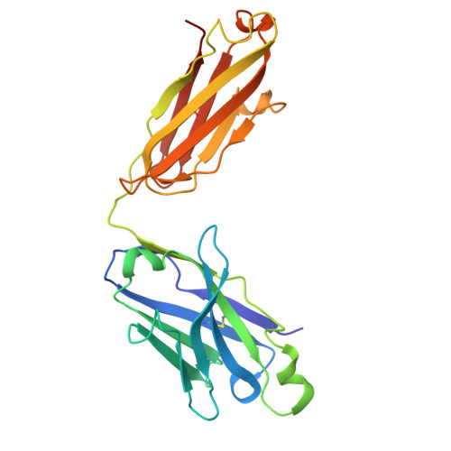
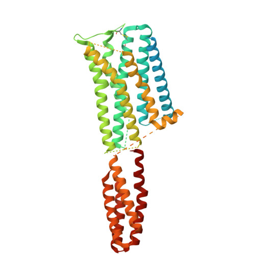
Inactive structures of the vasopressin V2 receptor reveal distinct binding modes for Tolvaptan and Mambaquaretin toxin.

Fouillen, A.Bous, J.Couvineau, P.Orcel, H.Mary, C.Lafleur, L.Pierre, T.Mendre, C.Gilles, N.Schulte, G.Granier, S.Mouillac, B.

(2025) Nat Commun 16: 3899-3899

  • DOI: https://doi.org/10.1038/s41467-025-59114-5
  • Primary Citation of Related Structures:  
    9HAP, 9HB3

  • PubMed Abstract: 

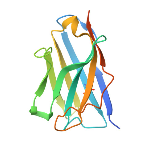
    Inhibitors of the arginine-vasopressin (AVP) V2 receptor (V2R) are key therapeutic compounds for treating hyponatremia or polycystic kidney diseases. Rational drug design based on experimental G protein-coupled receptor structures is a powerful avenue to develop better drugs. So far, the lack of inhibitor-bound V2R structures has impaired this strategy. Here we describe the cryo-electron microscopy structures of the V2R in complex with two selective inverse agonists, the non-peptide Tolvaptan (TVP) and the green mamba snake Mambaquaretin toxin (MQ1). Both ligands bind into the orthosteric binding site but with substantial differences. TVP binds deeper than MQ1, and directly contacts the toggle switch residue W284 6.48 in the transmembrane domain 6. The Kunitz-fold toxin displays extensive contacts with extracellular and transmembrane residues. As anticipated from TVP and MQ1 pharmacological properties, both structures represent inactive V2R conformations. Their comparison with those of the active AVP-bound V2R reveals the molecular mechanisms modulating receptor activity. The mini-protein MQ1-bound V2R structure suggests a new pharmacology approach for treating water homeostasis and renal diseases.


  • Organizational Affiliation

    Institut de Génomique Fonctionnelle, Université de Montpellier, CNRS, INSERM, Montpellier cedex 5, France.


Macromolecules
Find similar proteins by:  (by identity cutoff)  |  3D Structure
Entity ID: 1
MoleculeChains Sequence LengthOrganismDetailsImage
Synthetic antibody, anti-BRIL Fab fragment, Heavy chainA [auth H]231Homo sapiensMutation(s): 0 
Entity Groups  
Sequence Clusters30% Identity50% Identity70% Identity90% Identity95% Identity100% Identity
Sequence Annotations
Expand
  • Reference Sequence
Find similar proteins by:  (by identity cutoff)  |  3D Structure
Entity ID: 2
MoleculeChains Sequence LengthOrganismDetailsImage
anti-BRIL Fab NanobodyB [auth K]141Homo sapiensMutation(s): 0 
Entity Groups  
Sequence Clusters30% Identity50% Identity70% Identity90% Identity95% Identity100% Identity
Sequence Annotations
Expand
  • Reference Sequence
Find similar proteins by:  (by identity cutoff)  |  3D Structure
Entity ID: 3
MoleculeChains Sequence LengthOrganismDetailsImage
Synthetic antibody, anti-BRIL Fab fragment, Light chainC [auth L]215Homo sapiensMutation(s): 0 
Entity Groups  
Sequence Clusters30% Identity50% Identity70% Identity90% Identity95% Identity100% Identity
Sequence Annotations
Expand
  • Reference Sequence
Find similar proteins by:  (by identity cutoff)  |  3D Structure
Entity ID: 4
MoleculeChains Sequence LengthOrganismDetailsImage
Vasopressin V2 receptor,Soluble cytochrome b562D [auth R]552Homo sapiensEscherichia coli
This entity is chimeric
Mutation(s): 2 
Gene Names: AVPR2ADHRDIRDIR3V2RcybC
UniProt & NIH Common Fund Data Resources
Find proteins for P0ABE7 (Escherichia coli)
Explore P0ABE7 
Go to UniProtKB:  P0ABE7
Find proteins for P30518 (Homo sapiens)
Explore P30518 
Go to UniProtKB:  P30518
PHAROS:  P30518
GTEx:  ENSG00000126895 
Entity Groups  
Sequence Clusters30% Identity50% Identity70% Identity90% Identity95% Identity100% Identity
UniProt GroupsP0ABE7P30518
Sequence Annotations
Expand
  • Reference Sequence
Small Molecules
Ligands 1 Unique
IDChains Name / Formula / InChI Key2D Diagram3D Interactions
A1IT8 (Subject of Investigation/LOI)
Query on A1IT8

Download Ideal Coordinates CCD File 
E [auth R](R)-Tolvaptan
C26 H25 Cl N2 O3
GYHCTFXIZSNGJT-XMMPIXPASA-N
Experimental Data & Validation

Experimental Data

  • Method: ELECTRON MICROSCOPY
  • Resolution: 2.50 Å
  • Aggregation State: PARTICLE 
  • Reconstruction Method: SINGLE PARTICLE 
EM Software:
TaskSoftware PackageVersion
RECONSTRUCTIONcryoSPARC4.5.1
MODEL REFINEMENTRosetta
MODEL REFINEMENTPHENIX

Structure Validation

View Full Validation Report


Entry History & Funding Information

Deposition Data


Funding OrganizationLocationGrant Number
Agence Nationale de la Recherche (ANR)FranceANR-19-CE11-0014
Agence Nationale de la Recherche (ANR)FranceANR-22-CE44-0021
H2020 Marie Curie Actions of the European CommissionEuropean Union101063049_3D-V2R

Revision History  (Full details and data files)

  • Version 1.0: 2025-06-04
    Type: Initial release