


Crystal structure of Ni2+ dependent glycerol-1-phosphate dehydrogenase AraM from Bacillus subtilis
Mao, T., Pijning, T., Guskov, A.To be published.
Experimental Data Snapshot
Starting Model: in silico
View more details
Entity ID: 1 | |||||
|---|---|---|---|---|---|
| Molecule | Chains | Sequence Length | Organism | Details | Image |
| Glycerol-1-phosphate dehydrogenase [NAD(P)+] | 414 | Bacillus subtilis | Mutation(s): 0 Gene Names: egsA, araM, yseB, BSU28760 EC: 1.1.1.261 | 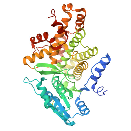 | |
UniProt | |||||
Entity Groups | |||||
| Sequence Clusters | 30% Identity50% Identity70% Identity90% Identity95% Identity100% Identity | ||||
| UniProt Group | P94527 | ||||
Sequence AnnotationsExpand | |||||
Reference Sequence | |||||
| Ligands 3 Unique | |||||
|---|---|---|---|---|---|
| ID | Chains | Name / Formula / InChI Key | 2D Diagram | 3D Interactions | |
| NAI (Subject of Investigation/LOI) Download:Ideal Coordinates CCD File | C [auth A] | 1,4-DIHYDRONICOTINAMIDE ADENINE DINUCLEOTIDE C21 H29 N7 O14 P2 BOPGDPNILDQYTO-NNYOXOHSSA-N |  | ||
| 13P (Subject of Investigation/LOI) Download:Ideal Coordinates CCD File | B [auth A] | 1,3-DIHYDROXYACETONEPHOSPHATE C3 H7 O6 P GNGACRATGGDKBX-UHFFFAOYSA-N |  | ||
| NI (Subject of Investigation/LOI) Download:Ideal Coordinates CCD File | D [auth A] | NICKEL (II) ION Ni VEQPNABPJHWNSG-UHFFFAOYSA-N |  | ||
| Length ( Å ) | Angle ( ˚ ) |
|---|---|
| a = 92.015 | α = 90 |
| b = 70.499 | β = 95.51 |
| c = 72.609 | γ = 90 |
| Software Name | Purpose |
|---|---|
| REFMAC | refinement |
| XDS | data reduction |
| Aimless | data scaling |
| PHASER | phasing |
| Funding Organization | Location | Grant Number |
|---|---|---|
| Not funded | -- |


