Biochemical and structural insights into pinoresinol hydroxylase from Pseudomonas sp.
Guerriere, T.B., Fraaije, M.W., Mattevi, A.(2024) Arch Biochem Biophys 764: 110247-110247
- PubMed: 39613284 Search on PubMed
- DOI: https://doi.org/10.1016/j.abb.2024.110247
- Primary Citation Related Structures:
9H40 - PubMed Abstract:
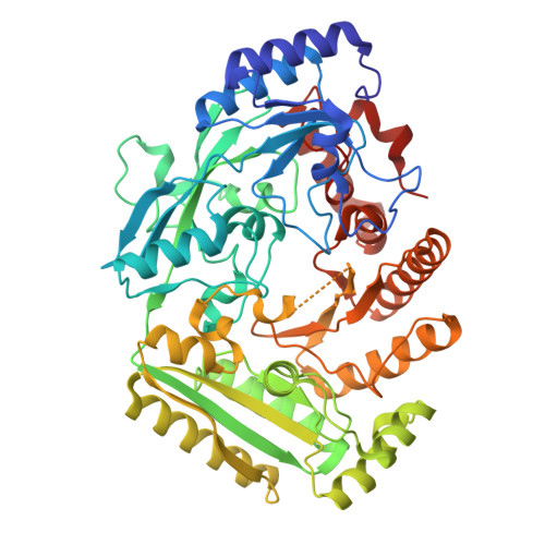
The vanillyl alcohol oxidase/p-cresol methylhydroxylase (VAO/PCMH) flavoprotein family comprises a broad spectrum of enzymes capable of catalyzing the oxidative bioconversions of various substrates. Among them, pinoresinol hydroxylase (PinH) from the 4-alkylphenol oxidizing subgroup initiates the oxidative degradation of (+)-pinoresinol, a lignan important for both lignin structure and plant defense. In this study, we present a detailed biochemical and structural characterization of PinH from Pseudomonas sp., with focus on its substrate specificity and product formation. PinH was expressed in E. coli and purified as FAD-containing, soluble protein. The flavoenzyme catalyzes the hydroxylation of both (+)-pinoresinol and eugenol. Structural analysis reveals its dimeric form, non-covalent flavin binding, and a large active site. AlphaFold models of the PinH-cytochrome complex demonstrate cytochrome's dual role in electron transfer and modulating PinH's conformation. A distinctive feature of PinH is a large cavity that hosts its multi-ring (+)-pinoresinol substrate. The capability of converting bulky lignans is particularly attractive for biotechnological applications aimed at producing high-value compounds from phenolic precursors. These insights expand our knowledge on the structure and mechanism of the VAO/PCMH flavoenzyme family members.
- Department of Biology and Biotechnology "Lazzaro Spallanzani", University of Pavia, Pavia, Italy.
Organizational Affiliation: