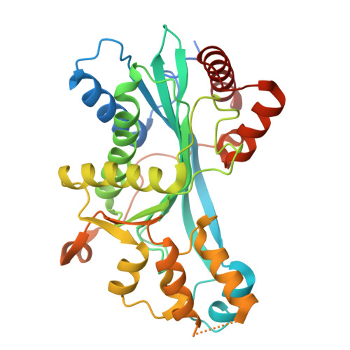
Structural and functional characterization of CREB-binding protein (CREBBP) as a histone propionyltransferase.
Cui, G., Ley, M., Mechaly, A.E., Bui, L.C., Michail, C., Berthelet, J., Dairou, J., Yang, H., Chevreux, G., Moroy, G., Green, M.R., Haouz, A., Rodrigues Lima, F.(2025) J Biological Chem 301: 110444-110444
- PubMed: 40615044 Search on PubMedSearch on PubMed Central
- DOI: https://doi.org/10.1016/j.jbc.2025.110444
- Primary Citation Related Structures:
9H02, 9H0K - PubMed Abstract:
In addition to histone acetylation, histone lysine propionylation (such as the H3K18Pr mark) has recently attracted significant attention as a common and abundant modification linking the cellular metabolic state and gene expression. CREB-binding protein (CREBBP) and EP300 are key histone acetyltransferases that play a critical role in gene expression through their catalytic activity. Although CREBBP and EP300 are homologous enzymes with high structural similarities, they exhibit both redundant and specific functions. Dissecting the shared and divergent properties of CREBBP and EP300 is thus important to understand their roles. However, despite the importance of CREBBP, most mechanistic and structural studies have focused on EP300, leaving much less information about CREBBP. Interestingly, recent enzymatic and structural studies have demonstrated that EP300 can also function as a histone propionyltransferase. Using a combination of acyltransferase assays with different acyl-CoA cofactors and with peptides, recombinant histone, or recombinant nucleosomes as substrates, we provide enzymatic evidence that, in addition to its well-documented acetyltransferase activity, CREBBP readily propionylates histone H3 in vitro, notably generating the H3K18Pr mark. Importantly, subsequent cellular studies using CRISPR-Cas9-edited cells further support that CREBBP can act as a histone propionyltransferase in vivo, notably depositing the H3K18Pr mark. Finally, the crystal structures of the human CREBBP histone acetyltransferase domain in complex with propionyl-CoA or in complex with Lys-CoA provide the structural basis for the histone propionyltransferase properties of CREBBP. Taken together, these findings provide new insights into the enzymatic functions of CREBBP and a better understanding of the mechanisms linking cellular metabolism and epigenetic regulation.
- Université Paris Cité, CNRS, Unité de Biologie Fonctionnelle et Adaptative, Paris, France.
Organizational Affiliation: