9GXZ | pdb_00009gxz

Structure of the Medicago truncatula LYR4 ectodomain


Experimental Data Snapshot

  • Method: X-RAY DIFFRACTION
  • Resolution: 1.50 Å
  • R-Value Free: 
    0.183 (Depositor), 0.183 (DCC) 
  • R-Value Work: 
    0.146 (Depositor), 0.146 (DCC) 
  • R-Value Observed: 
    0.148 (Depositor) 

Starting Model: experimental
View more details

wwPDB Validation 3D Report Full Report

Validation slider image for 9GXZ

Ligand Structure Quality Assessment 


This is version 1.0 of the entry. See complete history

Literature

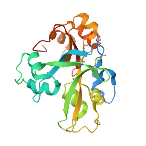
Structural basis for size-selective perception of chitin in plants

Gysel, K.Hansen, S.B.Ruebsam, H.Alsarraf, H.M.A.B.Madland, E.Cheng, J.X.J.Baadegaard, C.Poulsen, E.C.Vinther, M.Fort, S.Stougaard, J.Andersen, K.R.

To be published.

Macromolecule Content 

  • Total Structure Weight: 29.66 kDa 
  • Atom Count: 2,165 
  • Modeled Residue Count: 220 
  • Deposited Residue Count: 245 
  • Unique protein chains: 1

Macromolecules

Find similar proteins by:|  3D Structure
Entity ID: 1
MoleculeChains  Sequence LengthOrganismDetailsImage
LysM-domain receptor-like kinase245Medicago truncatulaMutation(s): 0 
Gene Names: 11433363MTR_5g085790MtrunA17_Chr5g0439331
UniProt
Find proteins for G7K7K0 (Medicago truncatula)
Explore G7K7K0 
Go to UniProtKB:  G7K7K0
Entity Groups
Sequence Clusters30% Identity50% Identity70% Identity90% Identity95% Identity100% Identity
UniProt GroupG7K7K0
Glycosylation
Glycosylation Sites: 6
Sequence Annotations
Expand
Reference Sequence

Small Molecules

Ligands 3 Unique
IDChains Name / Formula / InChI Key2D Diagram3D Interactions
NAG

Query on NAG


Download:Ideal Coordinates CCD File
B [auth A]
C [auth A]
D [auth A]
E [auth A]
F [auth A]
B [auth A],
C [auth A],
D [auth A],
E [auth A],
F [auth A],
G [auth A]
2-acetamido-2-deoxy-beta-D-glucopyranose
C8 H15 N O6
OVRNDRQMDRJTHS-FMDGEEDCSA-N
SO4

Query on SO4


Download:Ideal Coordinates CCD File
H [auth A],
I [auth A],
J [auth A],
K [auth A],
L [auth A]
SULFATE ION
O4 S
QAOWNCQODCNURD-UHFFFAOYSA-L
EDO

Query on EDO


Download:Ideal Coordinates CCD File
M [auth A]
N [auth A]
O [auth A]
P [auth A]
Q [auth A]
M [auth A],
N [auth A],
O [auth A],
P [auth A],
Q [auth A],
R [auth A],
S [auth A],
T [auth A],
U [auth A],
V [auth A]
1,2-ETHANEDIOL
C2 H6 O2
LYCAIKOWRPUZTN-UHFFFAOYSA-N
Modified Residues  1 Unique
IDChains TypeFormula2D DiagramParent
PCA
Query on PCA
A
L-PEPTIDE LINKINGC5 H7 N O3GLN

Experimental Data & Validation

Experimental Data

  • Method: X-RAY DIFFRACTION
  • Resolution: 1.50 Å
  • R-Value Free:  0.183 (Depositor), 0.183 (DCC) 
  • R-Value Work:  0.146 (Depositor), 0.146 (DCC) 
  • R-Value Observed: 0.148 (Depositor) 
Space Group: P 1 21 1
Unit Cell:
Length ( Å )Angle ( ˚ )
a = 32.247α = 90
b = 80.438β = 90.31
c = 46.898γ = 90
Software Package:
Software NamePurpose
PHENIXrefinement
PHENIXrefinement
XDSdata reduction
XDSdata scaling
PHASERphasing

Structure Validation

View Full Validation Report


Ligand Structure Quality Assessment 


Entry History 

& Funding Information

Deposition Data


Funding OrganizationLocationGrant Number
The Carlsberg FoundationDenmark--
Danish Council for Independent ResearchDenmark--
Novo Nordisk FoundationDenmark--

Revision History  (Full details and data files)

  • Version 1.0: 2025-07-16
    Type: Initial release