Gephyrin filaments represent the molecular basis of inhibitory postsynaptic densities.
Macha, A., Liebsch, F., Bruckisch, E.H.W., Burdina, N., von Stulpnagel, I., Benting, K., Gunkel, M., Behrmann, E., Schwarz, G.(2025) Nat Commun 16: 8293-8293
- PubMed: 40957893 Search on PubMedSearch on PubMed Central
- DOI: https://doi.org/10.1038/s41467-025-63748-w
- Primary Citation Related Structures:
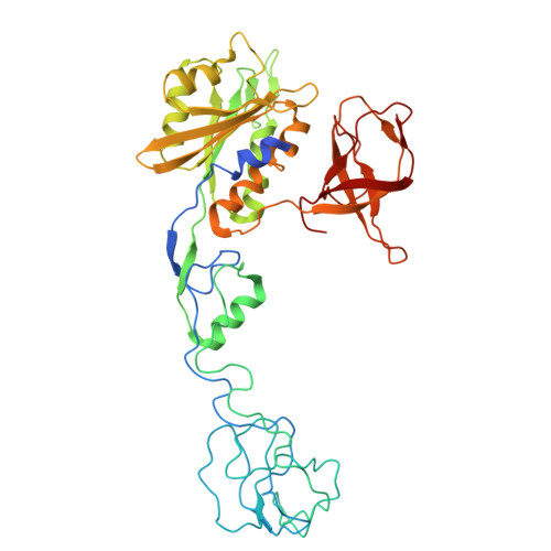
9GW9 - PubMed Abstract:
The multifunctional protein gephyrin clusters inhibitory receptors at the postsynaptic membrane in the CNS. Gephyrin has been proposed to form the inhibitory postsynaptic density by liquid-liquid phase separation, involving a complex interplay between receptor binding and oligomerization via its conserved G- and E-domains. Here we show by single particle cryo-EM analysis that dimerization promotes the formation of gephyrin filaments in which two E-domain dimers are linked by Z-shaped interfaces formed between two subdomains II (SDII) of adjacent dimers. Deletion of SDII, introduction of two epilepsy-causing pathogenic variants, or neutralization of an opposing charge in the interface abolish the formation of filaments, in vitro phase separation, and synaptic receptor clustering in hippocampal neurons. In conclusion, this work identifies gephyrin E-domain filaments as the structural foundation underlying gephyrin both phase separation and receptor clustering at inhibitory postsynaptic densities.
- Institute of Biochemistry, Department of Chemistry and Biochemistry, University of Cologne, Cologne, Germany.
Organizational Affiliation: