9GSN | pdb_00009gsn

Structure of the ATPgS-S1 state of the heptameric Bcs1 AAA-ATPase


Experimental Data Snapshot

  • Method: ELECTRON MICROSCOPY
  • Resolution: 2.58 Å
  • Aggregation State: PARTICLE 
  • Reconstruction Method: SINGLE PARTICLE 

wwPDB Validation 3D Report Full Report

Validation slider image for 9GSN

This is version 2.1 of the entry. See complete history

Literature

Mechanistic insights into Bcs1-mediated mitochondrial membrane translocation of the folded Rieske protein.

Rosales-Hernandez, C.Thoms, M.Berninghausen, O.Becker, T.Beckmann, R.

(2025) EMBO J 44: 3720-3741

  • DOI: https://doi.org/10.1038/s44318-025-00459-4
  • Primary Citation Related Structures: 
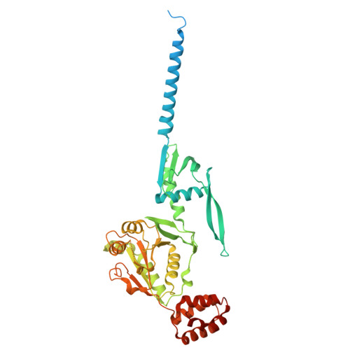
    9GS2, 9GSN, 9GU9

  • PubMed Abstract: 

    A functional mitochondrial respiratory chain requires coordinated and tightly regulated assembly of mitochondrial- and nuclear-encoded subunits. For bc1 complex (complex III) assembly, the iron-sulfur protein Rip1 must first be imported into the mitochondrial matrix to fold and acquire its 2Fe-2S cluster, then translocated and inserted into the inner mitochondrial membrane (IM). This translocation of folded Rip1 is accomplished by Bcs1, an unusual heptameric AAA ATPase that couples ATP hydrolysis to translocation. However, the molecular and mechanistic details of Bcs1-mediated Rip1 translocation have remained elusive. Here, we provide structural and biochemical evidence on how Bcs1 alternates between conformational states to translocate Rip1 across the IM. Using cryo-electron microscopy (cryo-EM), we identified substrate-bound pre-translocation and pre-release states, revealing how electrostatic interactions promote Rip1 binding to Bcs1. An ATP-induced conformational switch of the Bcs1 heptamer facilitates Rip1 translocation between two distinct aqueous vestibules-one exposed to the matrix, the other to the intermembrane space-in an airlock-like mechanism. This would minimize disruption of the IM permeability barrier, which could otherwise lead to proton leakage and compromised mitochondrial energy conversion.


  • Organizational Affiliation
    • Department of Biochemistry, Gene Center, University of Munich, Feodor-Lynen-Str. 25, 81377, Munich, Germany.

Macromolecule Content 

  • Total Structure Weight: 380.45 kDa 
  • Atom Count: 21,574 
  • Modeled Residue Count: 2,695 
  • Deposited Residue Count: 3,360 
  • Unique protein chains: 1

Macromolecules

Find similar proteins by:|  3D Structure
Entity ID: 1
MoleculeChains  Sequence LengthOrganismDetailsImage
Mitochondrial chaperone BCS1
A, B, C, D, E
A, B, C, D, E, F, G
480Saccharomyces cerevisiaeMutation(s): 0 
Gene Names: BCS1YDR375CD9481.17
EC: 3.6.1
UniProt
Find proteins for P32839 (Saccharomyces cerevisiae (strain ATCC 204508 / S288c))
Explore P32839 
Go to UniProtKB:  P32839
Entity Groups
Sequence Clusters30% Identity50% Identity70% Identity90% Identity95% Identity100% Identity
UniProt GroupP32839
Sequence Annotations
Expand
Reference Sequence

Experimental Data & Validation

Experimental Data

  • Method: ELECTRON MICROSCOPY
  • Resolution: 2.58 Å
  • Aggregation State: PARTICLE 
  • Reconstruction Method: SINGLE PARTICLE 
EM Software:
TaskSoftware PackageVersion
MODEL REFINEMENTPHENIX1.20.1_4487:

Structure Validation

View Full Validation Report


Entry History 

& Funding Information

Deposition Data


Funding OrganizationLocationGrant Number
German Research Foundation (DFG)Germany--

Revision History  (Full details and data files)

  • Version 1.0: 2025-06-04
    Type: Initial release
  • Version 2.0: 2025-06-11
    Type: Coordinate replacement
    Reason: Ligand geometry
    Changes: Advisory, Atomic model, Data collection, Derived calculations, Refinement description
  • Version 2.1: 2025-07-16
    Changes: Data collection, Database references