9GRF | pdb_00009grf

Crystal structure of X409 complexed to tetra-Tn-glycopeptide


Experimental Data Snapshot

  • Method: X-RAY DIFFRACTION
  • Resolution: 1.14 Å
  • R-Value Free: 
    0.172 (Depositor), 0.171 (DCC) 
  • R-Value Work: 
    0.156 (Depositor), 0.158 (DCC) 
  • R-Value Observed: 
    0.157 (Depositor) 

Starting Model: experimental
View more details

wwPDB Validation   3D Report Full Report


Ligand Structure Quality Assessment 


This is version 1.0 of the entry. See complete history


Literature

Microbial binding module employs sophisticated clustered saccharide patches to selectively adhere to mucins.

Jaroentomeechai, T.Veloz, B.Soares, C.O.Goerdeler, F.Grosso, A.S.Bull, C.Miller, R.L.Furukawa, S.Gines-Alcober, I.Taleb, V.Merino, P.Ghirardello, M.Companon, I.Coelho, H.Dias, J.S.Vincentelli, R.Henrissat, B.Joshi, H.Clausen, H.Corzana, F.Marcelo, F.Hurtado-Guerrero, R.Narimatsu, Y.

(2025) Nat Commun 16: 9058-9058

  • DOI: https://doi.org/10.1038/s41467-025-63756-w
  • Primary Citation of Related Structures:  
    9GRF, 9GRJ, 9GSM

  • PubMed Abstract: 

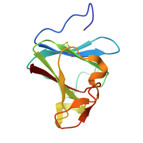
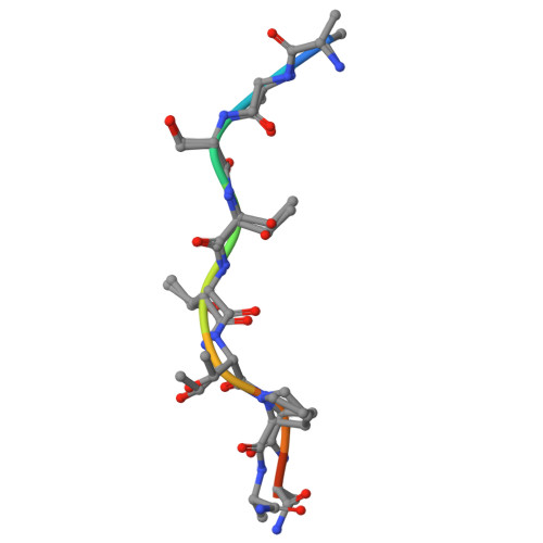
    The mucus lining wet body surfaces forms the interphase and barrier for the microbiota and resident microbiomes. Large mucin proteins densely decorated with O-glycans make up the mucus lining to entrap, feed and shape the microbiota, and repress biofilm formation and virulence. How mucins exert these effects is poorly understood and critical is how the microbiota recognize, sense, and break down mucins. Here, we provide structural molecular evidence that a small mucin-binding module designated X409 recognizes clustered saccharide patches comprised of rows of inner monosaccharides in adjacent O-glycans. These patches are unique to mucins and binding to these provides an elegant mechanism to retain adherence to mucins despite trimming of O-glycans during microbial scavenging of monosaccharides from mucins. Realization of clustered saccharide patch-binding motifs provides a hitherto overlooked scenario of contextual glycan epitopes and impetus for discovery of new classes of glycan-binding proteins.


  • Organizational Affiliation
    • Copenhagen Center for Glycomics, Departments of Cellular and Molecular Medicine, Faculty of Health Sciences, University of Copenhagen, Copenhagen, Denmark.

Macromolecules
Find similar proteins by:  (by identity cutoff)  |  3D Structure
Entity ID: 1
MoleculeChains Sequence LengthOrganismDetailsImage
Metalloprotease StcE104Escherichia coliMutation(s): 0 
Gene Names: stcEtagAL7031ECO57PM83
EC: 3.4.24
UniProt
Find proteins for O82882 (Escherichia coli O157:H7)
Explore O82882 
Go to UniProtKB:  O82882
Entity Groups  
Sequence Clusters30% Identity50% Identity70% Identity90% Identity95% Identity100% Identity
UniProt GroupO82882
Sequence Annotations
Expand
  • Reference Sequence

Find similar proteins by:  Sequence   |   3D Structure  

Entity ID: 2
MoleculeChains Sequence LengthOrganismDetailsImage
tetra-Tn-glycopeptideB [auth M]10synthetic constructMutation(s): 0 
Entity Groups  
Sequence Clusters30% Identity50% Identity70% Identity90% Identity95% Identity100% Identity
Glycosylation
Glycosylation Sites: 4
Sequence Annotations
Expand
  • Reference Sequence
Experimental Data & Validation

Experimental Data

  • Method: X-RAY DIFFRACTION
  • Resolution: 1.14 Å
  • R-Value Free:  0.172 (Depositor), 0.171 (DCC) 
  • R-Value Work:  0.156 (Depositor), 0.158 (DCC) 
  • R-Value Observed: 0.157 (Depositor) 
Space Group: P 21 21 2
Unit Cell:
Length ( Å )Angle ( ˚ )
a = 63.696α = 90
b = 76.05β = 90
c = 27.858γ = 90
Software Package:
Software NamePurpose
REFMACrefinement
XDSdata reduction
SCALAdata scaling
PHASERphasing

Structure Validation

View Full Validation Report


Ligand Structure Quality Assessment 


Entry History & Funding Information

Deposition Data


Funding OrganizationLocationGrant Number
Ministerio de Ciencia e Innovacion (MCIN)SpainPID2022-136362NB-I00

Revision History  (Full details and data files)

  • Version 1.0: 2025-12-24
    Type: Initial release