9GMQ | pdb_00009gmq

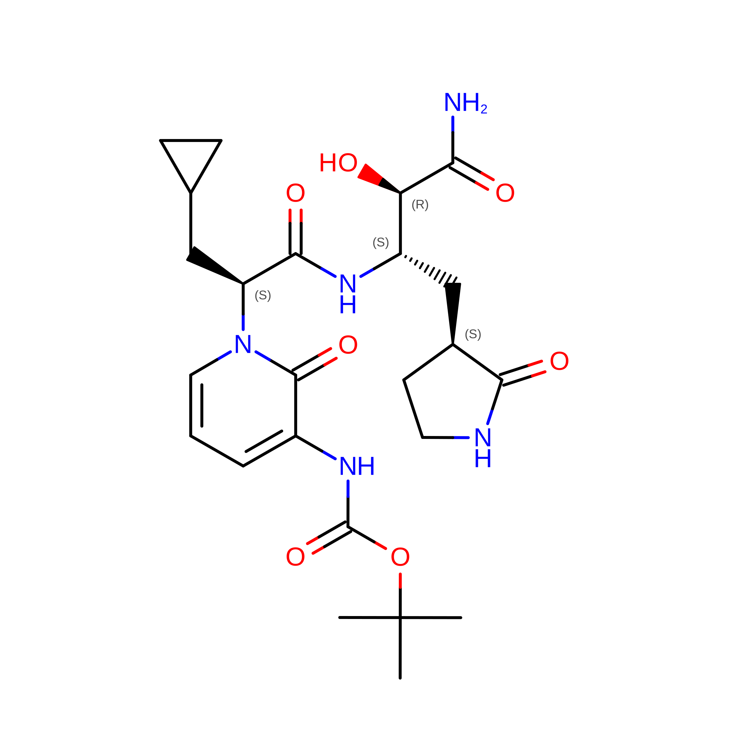
Crystal structure of the Mpro of SARS COV-2 in complex with the MG-87 inhibitor


Experimental Data Snapshot

  • Method: X-RAY DIFFRACTION
  • Resolution: 2.19 Å
  • R-Value Free: 
    0.218 (Depositor), 0.215 (DCC) 
  • R-Value Work: 
    0.166 (Depositor) 

Starting Model: experimental
View more details

wwPDB Validation 3D Report Full Report

Validation slider image for 9GMQ

Ligand Structure Quality Assessment 


This is version 2.1 of the entry. See complete history

Literature

Investigating the Binding Mode of a Naphthol-Based Inhibitor Targeting SARS-CoV-2 Main Protease.

Akrani, I.El Kilani, H.Touret, F.Kouvali, S.Gourni, V.Tsoka, A.Tsetis, J.K.Vorgias, C.Hilgenfeld, R.Kostakis, I.K.Myrianthopoulos, V.Mikros, E.

(2026) ChemMedChem 21: e70448-e70448

  • DOI: https://doi.org/10.1002/cmdc.70448
  • Primary Citation Related Structures: 
    32NU, 9GMQ

  • PubMed Abstract: 

    Regardless of the massive global efforts to combat the virus causing SARS-CoV-2 syndrome, the infection remains a substantial health challenge worldwide in the years following 2019. To this direction, targeting the main viral protease M pro has been proposed as a tractable and particularly promising approach toward developing effective and safe COVID-19 antivirals. By applying an integrated workflow combining a previously developed in silico consensus ranking protocol with two orthogonal in vitro methods, the NCI/DTP repository is screened and the discovery of an original naphthol scaffold with M pro inhibitory properties is reported. The hit is characterized in terms of structure and binding thermodynamics by combining X-ray crystallography and isothermal titration calorimetry where a binding affinity constant of 1.55 μM is determined. The compound is further evaluated against virus-infected cells, where an EC 50 value of 7.23 μM and comparable toxicity with nirmatrelvir are measured. Chemical synthesis is additionally employed to facilitate optimal exploration of the structure-activity relationship landscape regarding the new hit. By integrating computational, biophysical, and enzymatic methods, the suggested approach allows the combination of a structural hypothesis with functional evidence and shows its capacity toward identifying and rationally optimizing structurally original noncovalent M pro inhibitors.


  • Organizational Affiliation
    • Department of Pharmacy, National and Kapodistrian University of Athens, Athens, Greece.

Macromolecule Content 

  • Total Structure Weight: 68.7 kDa 
  • Atom Count: 4,988 
  • Modeled Residue Count: 612 
  • Deposited Residue Count: 612 
  • Unique protein chains: 1

Macromolecules

Find similar proteins by:|  3D Structure
Entity ID: 1
MoleculeChains  Sequence LengthOrganismDetailsImage
3C-like proteinase nsp5
A, B
306Severe acute respiratory syndrome coronavirus 2Mutation(s): 0 
EC: 3.4.22.69
UniProt
Find proteins for P0DTC1 (Severe acute respiratory syndrome coronavirus 2)
Explore P0DTC1 
Go to UniProtKB:  P0DTC1
Entity Groups
Sequence Clusters30% Identity50% Identity70% Identity90% Identity95% Identity100% Identity
UniProt GroupP0DTC1
Sequence Annotations
Expand
Reference Sequence

Small Molecules

Ligands 2 Unique
IDChains Name / Formula / InChI Key2D Diagram3D Interactions
MXU
(Subject of Investigation/LOI)

Query on MXU


Download:Ideal Coordinates CCD File
C [auth A],
E [auth B]
~{tert}-butyl ~{N}-[1-[(2~{S})-1-[[(2~{S},3~{R})-4-azanyl-3-oxidanyl-4-oxidanylidene-1-[(3~{S})-2-oxidanylidenepyrrolidin-3-yl]butan-2-yl]amino]-3-cyclopropyl-1-oxidanylidene-propan-2-yl]-2-oxidanylidene-pyridin-3-yl]carbamate
C24 H35 N5 O7
WNCHATFCLSCVLN-LEUOFYLZSA-N
CL

Query on CL


Download:Ideal Coordinates CCD File
D [auth A]CHLORIDE ION
Cl
VEXZGXHMUGYJMC-UHFFFAOYSA-M

Experimental Data & Validation

Experimental Data

  • Method: X-RAY DIFFRACTION
  • Resolution: 2.19 Å
  • R-Value Free:  0.218 (Depositor), 0.215 (DCC) 
  • R-Value Work:  0.166 (Depositor) 
Space Group: P 1 21 1
Unit Cell:
Length ( Å )Angle ( ˚ )
a = 45.941α = 90
b = 63.773β = 91.251
c = 103.513γ = 90
Software Package:
Software NamePurpose
REFMACrefinement
XDSdata reduction
Aimlessdata scaling
MOLREPphasing

Structure Validation

View Full Validation Report


Ligand Structure Quality Assessment 


Entry History 

& Funding Information

Deposition Data


Funding OrganizationLocationGrant Number
European CommissionEuropean Union--

Revision History  (Full details and data files)

  • Version 1.0: 2025-01-29
    Type: Initial release
  • Version 2.0: 2025-02-26
    Changes: Atomic model, Data collection, Database references
  • Version 2.1: 2026-09-02
    Changes: Database references