9GL5 | pdb_00009gl5

X-ray structure of the Thermus thermophilus Q190E mutant of the PilF-GSPIIB domain in the c-di-GMP bound state


Experimental Data Snapshot

  • Method: X-RAY DIFFRACTION
  • Resolution: 1.90 Å
  • R-Value Free: 
    0.225 (Depositor), 0.227 (DCC) 
  • R-Value Work: 
    0.193 (Depositor), 0.194 (DCC) 
  • R-Value Observed: 
    0.194 (Depositor) 

Starting Model: experimental
View more details

wwPDB Validation 3D Report Full Report

Validation slider image for 9GL5

Ligand Structure Quality Assessment 


This is version 1.1 of the entry. See complete history

Literature

Protonated Glutamate and Aspartate Side Chains Can Recognize Phosphodiester Groups via Strong and Short Hydrogen Bonds in Biomacromolecular Complexes.

Neissner, K.Duchardt-Ferner, E.Wiedemann, C.Kraus, J.Hellmich, U.A.Wohnert, J.

(2025) Angew Chem Int Ed Engl 64: e202501589-e202501589

  • DOI: https://doi.org/10.1002/anie.202501589
  • Primary Citation Related Structures: 
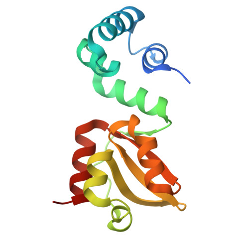
    9GL5, 9GLG

  • PubMed Abstract: 

    Phosphodiester groups occur ubiquitously in nature, e.g. in nucleic acids or in cyclic (di-)nucleotides important for signal transduction. Proteins often use polar or positively charged amino acids to interact with the negatively charged phosphodiester groups via hydrogen bonds and salt bridges. In contrast, the acidic amino acids aspartate and glutamate are generally not considered as important determinants for phosphodiester group recognition. Instead, they are regarded as detrimental to such interactions due to the assumed charge repulsion between their deprotonated, negatively charged side chain carboxylate groups and the phosphodiester. Accordingly, acidic amino acids are often purposefully introduced into proteins to abrogate nucleic acid interactions in functional studies. Here, we show that in appropriate structural contexts, glutamate side chains are readily protonated even at neutral pH and act as hydrogen bond donors to phosphodiester groups using a c-di-GMP binding protein - the GSPII-B domain of PilF from Thermus thermophilus - as an example. Surveying available RNA-protein and DNA-protein complex structures in the PDB, we found that hydrogen bonds between apparently protonated carboxylate groups of glutamate and aspartate and phosphodiester groups occur frequently in many different functional protein classes. Thus, the functional role of acidic amino acids in phosphodiester group recognition needs to be re-evaluated.


  • Organizational Affiliation
    • Institute for Molecular Biosciences, Goethe-University Frankfurt/M., Max-von-Laue-Str. 9, 60438, Frankfurt, Germany.

Macromolecule Content 

  • Total Structure Weight: 34.74 kDa 
  • Atom Count: 2,376 
  • Modeled Residue Count: 274 
  • Deposited Residue Count: 292 
  • Unique protein chains: 1

Macromolecules

Find similar proteins by:|  3D Structure
Entity ID: 1
MoleculeChains  Sequence LengthOrganismDetailsImage
ATP-binding motif-containing protein pilF
A, B
146Thermus thermophilus HB27Mutation(s): 1 
Gene Names: pilFTT_C1622
UniProt
Find proteins for Q72H73 (Thermus thermophilus (strain ATCC BAA-163 / DSM 7039 / HB27))
Explore Q72H73 
Go to UniProtKB:  Q72H73
Entity Groups
Sequence Clusters30% Identity50% Identity70% Identity90% Identity95% Identity100% Identity
UniProt GroupQ72H73
Sequence Annotations
Expand
Reference Sequence

Small Molecules

Ligands 3 Unique
IDChains Name / Formula / InChI Key2D Diagram3D Interactions
C2E
(Subject of Investigation/LOI)

Query on C2E


Download:Ideal Coordinates CCD File
C [auth A],
H [auth B]
9,9'-[(2R,3R,3aS,5S,7aR,9R,10R,10aS,12S,14aR)-3,5,10,12-tetrahydroxy-5,12-dioxidooctahydro-2H,7H-difuro[3,2-d:3',2'-j][1,3,7,9,2,8]tetraoxadiphosphacyclododecine-2,9-diyl]bis(2-amino-1,9-dihydro-6H-purin-6-one)
C20 H24 N10 O14 P2
PKFDLKSEZWEFGL-MHARETSRSA-N
SO4

Query on SO4


Download:Ideal Coordinates CCD File
D [auth A]
E [auth A]
I [auth B]
J [auth B]
K [auth B]
D [auth A],
E [auth A],
I [auth B],
J [auth B],
K [auth B],
L [auth B]
SULFATE ION
O4 S
QAOWNCQODCNURD-UHFFFAOYSA-L
ACY

Query on ACY


Download:Ideal Coordinates CCD File
F [auth A],
G [auth A],
M [auth B],
N [auth B]
ACETIC ACID
C2 H4 O2
QTBSBXVTEAMEQO-UHFFFAOYSA-N

Experimental Data & Validation

Experimental Data

  • Method: X-RAY DIFFRACTION
  • Resolution: 1.90 Å
  • R-Value Free:  0.225 (Depositor), 0.227 (DCC) 
  • R-Value Work:  0.193 (Depositor), 0.194 (DCC) 
  • R-Value Observed: 0.194 (Depositor) 
Space Group: H 3
Unit Cell:
Length ( Å )Angle ( ˚ )
a = 107.966α = 90
b = 107.966β = 90
c = 86.366γ = 120
Software Package:
Software NamePurpose
MxCuBEdata collection
autoPROCdata processing
PHENIXphasing
autoPROCdata scaling
Cootmodel building
PHENIXrefinement
XDSdata scaling
XDSdata reduction

Structure Validation

View Full Validation Report


Ligand Structure Quality Assessment 


Entry History 

& Funding Information

Deposition Data


Funding OrganizationLocationGrant Number
German Research Foundation (DFG)Germany--

Revision History  (Full details and data files)

  • Version 1.0: 2025-06-25
    Type: Initial release
  • Version 1.1: 2025-07-09
    Changes: Database references