


Unraveling the Catalytic and Structural Properties of a New Bacterial Galactose Oxidase
Taborda, A., Frazao, T., Frazao, C., Borges, P.T., Martins, L.To be published.
Experimental Data Snapshot
Starting Model: in silico
View more details
Entity ID: 1 | |||||
|---|---|---|---|---|---|
| Molecule | Chains | Sequence Length | Organism | Details | Image |
| Galactose oxidase | 617 | Pseudarthrobacter siccitolerans | Mutation(s): 0 Gene Names: GAOA, ARTSIC4J27_1011 EC: 1.1.3.9 | 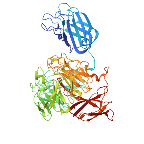 | |
UniProt | |||||
Find proteins for A0A024GZ97 (Pseudarthrobacter siccitolerans) Explore A0A024GZ97 Go to UniProtKB: A0A024GZ97 | |||||
Entity Groups | |||||
| Sequence Clusters | 30% Identity50% Identity70% Identity90% Identity95% Identity100% Identity | ||||
| UniProt Group | A0A024GZ97 | ||||
Sequence AnnotationsExpand | |||||
Reference Sequence | |||||
Entity ID: 2 | |||||
|---|---|---|---|---|---|
| Molecule | Chains | Sequence Length | Organism | Details | Image |
| SER-HIS-SER-SER-GLY-ALA | B [auth Q] | 6 | Pseudarthrobacter siccitolerans | Mutation(s): 0 |  |
| Ligands 6 Unique | |||||
|---|---|---|---|---|---|
| ID | Chains | Name / Formula / InChI Key | 2D Diagram | 3D Interactions | |
| PG4 (Subject of Investigation/LOI) Download:Ideal Coordinates CCD File | D [auth A] | TETRAETHYLENE GLYCOL C8 H18 O5 UWHCKJMYHZGTIT-UHFFFAOYSA-N |  | ||
| GAL (Subject of Investigation/LOI) Download:Ideal Coordinates CCD File | C [auth A] | beta-D-galactopyranose C6 H12 O6 WQZGKKKJIJFFOK-FPRJBGLDSA-N |  | ||
| SO4 (Subject of Investigation/LOI) Download:Ideal Coordinates CCD File | H [auth A] | SULFATE ION O4 S QAOWNCQODCNURD-UHFFFAOYSA-L |  | ||
| PO4 (Subject of Investigation/LOI) Download:Ideal Coordinates CCD File | F [auth A] | PHOSPHATE ION O4 P NBIIXXVUZAFLBC-UHFFFAOYSA-K |  | ||
| GOL (Subject of Investigation/LOI) Download:Ideal Coordinates CCD File | E [auth A] | GLYCEROL C3 H8 O3 PEDCQBHIVMGVHV-UHFFFAOYSA-N |  | ||
| CA (Subject of Investigation/LOI) Download:Ideal Coordinates CCD File | G [auth A] | CALCIUM ION Ca BHPQYMZQTOCNFJ-UHFFFAOYSA-N |  | ||
| Length ( Å ) | Angle ( ˚ ) |
|---|---|
| a = 48.005 | α = 90 |
| b = 61.406 | β = 90 |
| c = 186.396 | γ = 90 |
| Software Name | Purpose |
|---|---|
| REFMAC | refinement |
| XDS | data scaling |
| XDS | data reduction |
| PHASER | phasing |
| Funding Organization | Location | Grant Number |
|---|---|---|
| Foundation for Science and Technology (FCT) | Portugal | -- |


