9G0F | pdb_00009g0f

CryoEM structure of PmcTnsC-dsDNA-AMPPNP


Experimental Data Snapshot

  • Method: ELECTRON MICROSCOPY
  • Resolution: 3.70 Å
  • Aggregation State: PARTICLE 
  • Reconstruction Method: SINGLE PARTICLE 

wwPDB Validation 3D Report Full Report

Validation slider image for 9G0F

This is version 1.1 of the entry. See complete history

Literature

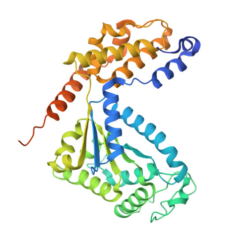
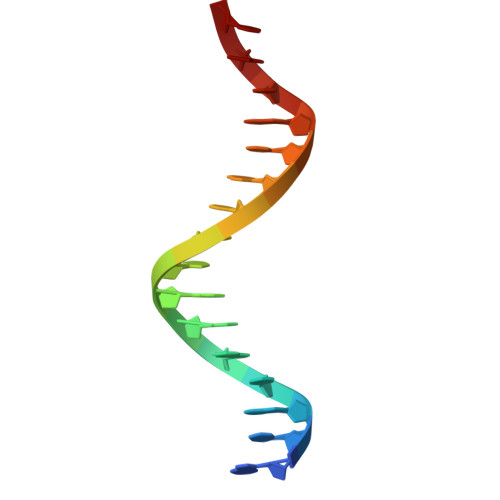
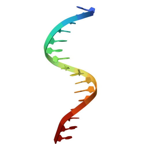
Structural basis of TnsC oligomerization and transposase recruitment in type I-B CRISPR-associated transposons.

Finocchio, G.Querques, I.Chanez, C.Speichert, K.J.Jinek, M.

(2025) Nucleic Acids Res 53

  • DOI: https://doi.org/10.1093/nar/gkaf149
  • Primary Citation Related Structures: 
    9G0F, 9GMZ

  • PubMed Abstract: 

    CRISPR-associated transposon (CAST) systems employ CRISPR-Cas systems as RNA-directed targeting modules for site-specific transposon DNA insertion. Among them, type I CASTs rely on the coordinated action of the guide RNA-bound Cascade complex and the transposon proteins TniQ, TnsC, and TnsAB. The interaction between the transposase TnsAB and the ATPase TnsC is crucial for transposition activity, yet the underlying molecular details have remained elusive. Here, we investigate the type I-B CAST system from Peltigera membranacea cyanobiont. Cryo-electron microscopic structures of TnsC and its complex with the C-terminal region of TnsAB reveal that TnsC forms a heptameric ring that recruits TnsAB by interacting with its C-terminal tail. In vitro binding assays indicate that TnsAB exclusively interacts with the TnsC heptamer without inducing its disassembly, in contrast to type V-K CAST systems. Mutational analysis of key structural features corroborates the significance of TnsC multimerization and TnsB interaction for transposon activity in vivo. Altogether, these findings offer detailed structural and functional insights into the molecular mechanism of type I-B CAST, with the aim of facilitating their development as genome engineering tools.


  • Organizational Affiliation
    • Department of Biochemistry, University of Zurich, 8057 Zurich, Switzerland.

Macromolecule Content 

  • Total Structure Weight: 320.07 kDa 
  • Atom Count: 20,077 
  • Modeled Residue Count: 2,421 
  • Deposited Residue Count: 2,734 
  • Unique protein chains: 1
  • Unique nucleic acid chains: 2

Macromolecules


Find similar proteins by:|  3D Structure
Entity ID: 1
MoleculeChains  Sequence LengthOrganismDetailsImage
AAA+ ATPase domain-containing protein
A, B, C, D, E
A, B, C, D, E, F, G
386Peltigera membranaceaMutation(s): 0 
Gene Names: CDG76_08990
UniProt
Find proteins for A0ACD6BA40 (Nostoc sp. 'Peltigera membranacea cyanobiont' 210A)
Explore A0ACD6BA40 
Go to UniProtKB:  A0ACD6BA40
Entity Groups
Sequence Clusters30% Identity50% Identity70% Identity90% Identity95% Identity100% Identity
UniProt GroupA0ACD6BA40
Sequence Annotations
Expand
Reference Sequence
Find similar nucleic acids by:  Sequence
Entity ID: 2
MoleculeChains LengthOrganismImage
DNAH [auth 1]16Escherichia coli
Sequence Annotations
Expand
Reference Sequence
Find similar nucleic acids by:  Sequence
Entity ID: 3
MoleculeChains LengthOrganismImage
DNAI [auth 2]16Escherichia coli
Sequence Annotations
Expand
Reference Sequence

Small Molecules

Ligands 2 Unique
IDChains Name / Formula / InChI Key2D Diagram3D Interactions
ANP
(Subject of Investigation/LOI)

Query on ANP


Download:Ideal Coordinates CCD File
J [auth A]
L [auth B]
N [auth C]
P [auth D]
R [auth E]
J [auth A],
L [auth B],
N [auth C],
P [auth D],
R [auth E],
T [auth F],
V [auth G]
PHOSPHOAMINOPHOSPHONIC ACID-ADENYLATE ESTER
C10 H17 N6 O12 P3
PVKSNHVPLWYQGJ-KQYNXXCUSA-N
MG
(Subject of Investigation/LOI)

Query on MG


Download:Ideal Coordinates CCD File
K [auth A]
M [auth B]
O [auth C]
Q [auth D]
S [auth E]
K [auth A],
M [auth B],
O [auth C],
Q [auth D],
S [auth E],
U [auth F],
W [auth G]
MAGNESIUM ION
Mg
JLVVSXFLKOJNIY-UHFFFAOYSA-N

Experimental Data & Validation

Experimental Data

  • Method: ELECTRON MICROSCOPY
  • Resolution: 3.70 Å
  • Aggregation State: PARTICLE 
  • Reconstruction Method: SINGLE PARTICLE 
EM Software:
TaskSoftware PackageVersion
MODEL REFINEMENTPHENIX

Structure Validation

View Full Validation Report


Entry History 

& Funding Information

Deposition Data


Funding OrganizationLocationGrant Number
European Research Council (ERC)European Union--

Revision History  (Full details and data files)

  • Version 1.0: 2025-04-02
    Type: Initial release
  • Version 1.1: 2025-07-09
    Changes: Data collection