9FXR | pdb_00009fxr

Crystal structure of trans-o-hydroxybenzylidenepyruvate hydratase-aldolase from Pseudomonas fluorescens N3 bound to pyruvate


Experimental Data Snapshot

  • Method: X-RAY DIFFRACTION
  • Resolution: 2.30 Å
  • R-Value Free: 
    0.226 (Depositor), 0.230 (DCC) 
  • R-Value Work: 
    0.200 (Depositor), 0.205 (DCC) 

Starting Model: experimental
View more details

wwPDB Validation 3D Report Full Report

Validation slider image for 9FXR

This is version 1.0 of the entry. See complete history

Literature

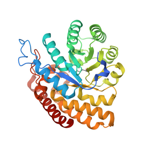
Structural snapshots of the aldol condensation reaction of the enzyme trans-o-hydroxybenzylidenepyruvate hydratase-aldolase from Pseudomonas fluorescens N3.

Ferrara, S.Braggiotti, B.Mastrangelo, E.Di Gennaro, P.Bertoni, G.Milani, M.

(2025) Biochem Biophys Res Commun 747: 151281-151281

  • DOI: https://doi.org/10.1016/j.bbrc.2024.151281
  • Primary Citation Related Structures: 
    9FRT, 9FTK, 9FXR

  • PubMed Abstract: 

    Aldolases are crucial enzymes that catalyze the formation of carbon-carbon bonds in the context of the anabolic and catabolic pathways of various metabolites. The bacterium Pseudomonas fluorescens N3 can use naphthalene as its sole carbon and energy source by using, among other enzymes, the trans-o-hydroxybenzylidenepyruvate (tHBP) hydratase-aldolase (HA), encoded by the nahE gene. In this study, we present the crystallographic structures of tHBP-HA in three different functional states: the apo enzyme with a phosphate ion in the active site, and the Schiff base adduct bound either to pyruvate or to the substitute with '(R)-4-hydroxy-4-(2-hydroxyphenyl)-2-oxobutanoate'(intermediate state). Our structures elucidate some of the phases of the aldol condensation reaction, proposing the role of a conserved water molecule (W2) in the deprotonation of the catalytic lysine. Moreover, our crystallographic data suggest potential pathways for substrate and product diffusion to and from the protein's active site. These insights advance our understanding of the molecular mechanisms of the aldolase function and can also be used for the design and optimization of new enzymes engineered for the chemical synthesis of different C-C adducts.


  • Organizational Affiliation
    • Biophysics Institute, CNR-IBF, Via Corti 12, I-20133, Milano, Italy; Department of Bioscience, University of Milan, Via Celoria 26, I-20133, Milano, Italy.

Macromolecule Content 

  • Total Structure Weight: 309.76 kDa 
  • Atom Count: 22,153 
  • Modeled Residue Count: 2,604 
  • Deposited Residue Count: 2,768 
  • Unique protein chains: 1

Macromolecules

Find similar proteins by:|  3D Structure
Entity ID: 1
MoleculeChains  Sequence LengthOrganismDetailsImage
Trans-O-hydroxybenzylidenepyruvate hydratase-aldolase346Pseudomonas fluorescensMutation(s): 0 
Gene Names: nahE
EC: 4.1.2.45
UniProt
Find proteins for C3KFM9 (Pseudomonas fluorescens)
Explore C3KFM9 
Go to UniProtKB:  C3KFM9
Entity Groups
Sequence Clusters30% Identity50% Identity70% Identity90% Identity95% Identity100% Identity
UniProt GroupC3KFM9
Sequence Annotations
Expand
Reference Sequence

Small Molecules

Ligands 3 Unique
IDChains Name / Formula / InChI Key2D Diagram3D Interactions
PO4

Query on PO4


Download:Ideal Coordinates CCD File
AA [auth L]
BA [auth L]
J [auth A]
M [auth B]
P [auth C]
AA [auth L],
BA [auth L],
J [auth A],
M [auth B],
P [auth C],
U [auth H],
V [auth H]
PHOSPHATE ION
O4 P
NBIIXXVUZAFLBC-UHFFFAOYSA-K
GOL

Query on GOL


Download:Ideal Coordinates CCD File
CA [auth L]
EA [auth N]
K [auth A]
N [auth B]
Q [auth C]
CA [auth L],
EA [auth N],
K [auth A],
N [auth B],
Q [auth C],
S [auth D],
W [auth H],
Y [auth J]
GLYCEROL
C3 H8 O3
PEDCQBHIVMGVHV-UHFFFAOYSA-N
PYR

Query on PYR


Download:Ideal Coordinates CCD File
DA [auth N]
I [auth A]
L [auth B]
O [auth C]
R [auth D]
DA [auth N],
I [auth A],
L [auth B],
O [auth C],
R [auth D],
T [auth H],
X [auth J],
Z [auth L]
PYRUVIC ACID
C3 H4 O3
LCTONWCANYUPML-UHFFFAOYSA-N
Modified Residues  1 Unique
IDChains TypeFormula2D DiagramParent
KPI
Query on KPI
A
B
C
D
E [auth H]
A,
B,
C,
D,
E [auth H],
F [auth J],
G [auth L],
H [auth N]
L-PEPTIDE LINKINGC9 H16 N2 O4LYS

Experimental Data & Validation

Experimental Data

  • Method: X-RAY DIFFRACTION
  • Resolution: 2.30 Å
  • R-Value Free:  0.226 (Depositor), 0.230 (DCC) 
  • R-Value Work:  0.200 (Depositor), 0.205 (DCC) 
Space Group: C 1 2 1
Unit Cell:
Length ( Å )Angle ( ˚ )
a = 200.172α = 90
b = 200.362β = 133.815
c = 144.473γ = 90
Software Package:
Software NamePurpose
REFMACrefinement
autoPROCdata reduction
autoPROCdata scaling
MOLREPphasing

Structure Validation

View Full Validation Report


Entry History 

& Funding Information

Deposition Data


Funding OrganizationLocationGrant Number
Not funded--

Revision History  (Full details and data files)

  • Version 1.0: 2025-07-02
    Type: Initial release