


Structural Characterization of B-DNA d(CGTGAATTCACG)2 in Complex with the Specific Minor Groove Binding Fluorescent Marker Hoechst 33342
Sbirkova-Dimitrova, H., Rusew, R., Gerginov, H., Heroux, A., Shivachev, B.L.(2025) Crystals (Basel) 15
Experimental Data Snapshot
Starting Model: experimental
View more details
(2025) Crystals (Basel) 15
Entity ID: 1 | ||||
| Molecule | Chains | Length | Organism | Image |
|---|---|---|---|---|
| DNA (5'-D(*CP*GP*TP*GP*AP*AP*TP*TP*CP*AP*CP*G)-3') | A [auth AAA], B [auth BBB] | 12 | DNA molecule | 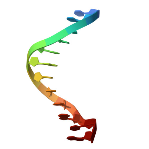 |
Sequence AnnotationsExpand | ||||
Reference Sequence | ||||
| Ligands 3 Unique | |||||
|---|---|---|---|---|---|
| ID | Chains | Name / Formula / InChI Key | 2D Diagram | 3D Interactions | |
| HT1 (Subject of Investigation/LOI) Download:Ideal Coordinates CCD File | G [auth BBB] | 2'-(4-ETHOXYPHENYL)-5-(4-METHYL-1-PIPERAZINYL)-2,5'-BI-BENZIMIDAZOLE C27 H28 N6 O PRDFBSVERLRRMY-UHFFFAOYSA-N |  | ||
| CL Download:Ideal Coordinates CCD File | E [auth AAA], F [auth AAA] | CHLORIDE ION Cl VEXZGXHMUGYJMC-UHFFFAOYSA-M |  | ||
| MG Download:Ideal Coordinates CCD File | C [auth AAA], D [auth AAA] | MAGNESIUM ION Mg JLVVSXFLKOJNIY-UHFFFAOYSA-N |  | ||
| Length ( Å ) | Angle ( ˚ ) |
|---|---|
| a = 24.593 | α = 90 |
| b = 40.406 | β = 90 |
| c = 65.033 | γ = 90 |
| Software Name | Purpose |
|---|---|
| REFMAC | refinement |
| XDS | data reduction |
| XSCALE | data scaling |
| PHASER | phasing |
| Funding Organization | Location | Grant Number |
|---|---|---|
| Bulgarian National Science Fund | Bulgaria | KP-06-KOCT/5 |


