Structural insight into the function of human peptidyl arginine deiminase 6.
Williams, J.P.C., Mouilleron, S., Trapero, R.H., Bertran, M.T., Marsh, J.A., Walport, L.J.(2024) Comput Struct Biotechnol J 23: 3258-3269
- PubMed: 39286527 Search on PubMedSearch on PubMed Central
- DOI: https://doi.org/10.1016/j.csbj.2024.08.019
- Primary Citation Related Structures:
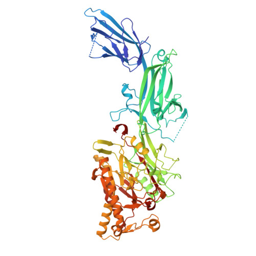
9FMN - PubMed Abstract:
Peptidyl arginine deiminase 6 (PADI6 or PAD6) is vital for early embryonic development in mice and humans, yet its function remains elusive. PADI6 is less conserved than other PADIs and it is currently unknown whether it has a catalytic function. Here we show that human PADI6 dimerises like hPADIs 2-4, however, does not bind Ca 2+ and is inactive in in vitro assays against standard PADI substrates. By determining the crystal structure of hPADI6, we show that hPADI6 is structured in the absence of Ca 2+ where hPADI2 and hPADI4 are not, and the Ca-binding sites are not conserved. Moreover, we show that whilst the key catalytic aspartic acid and histidine residues are structurally conserved, the cysteine is displaced far from the active site centre and the hPADI6 active site pocket appears closed through a unique evolved mechanism in hPADI6, not present in the other PADIs. Taken together, these findings provide insight into how the function of hPADI6 may differ from the other PADIs based on its structure and provides a resource for characterising the damaging effect of clinically significant PADI6 variants.
- Department of Chemistry, Imperial College London, London, United Kingdom.
Organizational Affiliation: